Tuesday, December 11, 2012

Light Microscopes

Light microscopes or thoughprovoking field microscopes are the most basic form of microscopes. The primary light microscope was invented way back in the 16th century. They are so called because they use light in the illustrated spectrum to illuminate the specimen. Light microscopes are extensively used everywhere from high-tech laboratories to schools.

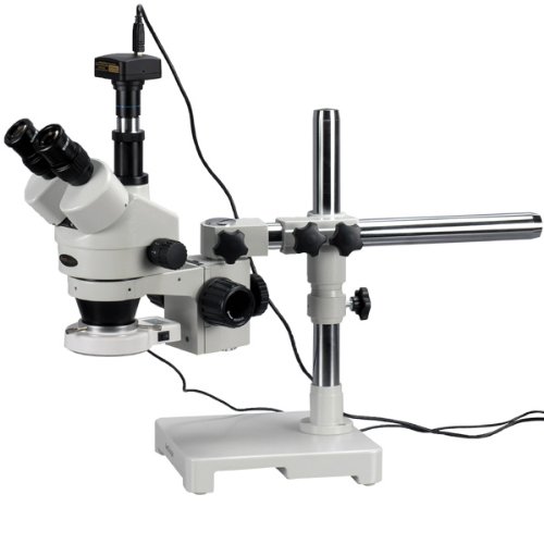
There are many types of light microscopes ready from just to 00 or even more. The major types of light microscopes are stereo or dissecting, easy and composition microscopes. Stereo microscopes are the most advanced and use two light paths for three-dimensional (3D) viewing. They are very costly and are used in laboratories and medicinal practices. easy light microscopes are used only for small scale magnification and they have only one lens both as eyepiece and objective.

Microscope

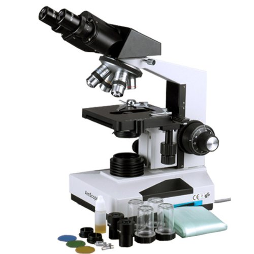
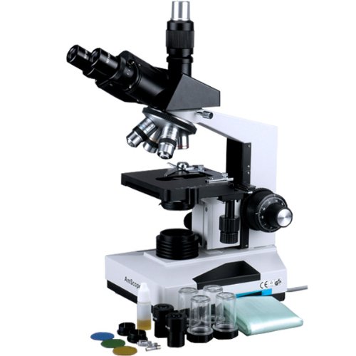
Compound microscopes are the most extensively used light microscopes. There are generally three grades of composition microscopes - student, benchtop and investigate light microscopes. Pupil microscopes are cheap, small and designed for thoughprovoking field, dark field, and phase disagreement examinations. Benchtop microscopes are used for test techniques. investigate microscopes are for investigate practices; they often contain built-in computers and cameras.

The components of a tasteless composition microscope are: a mirror or light source for reflecting or producing light, a condenser with diaphragms and filters to adjust the intensity of the light, a stage for supporting the specimen which contains a central hole to pass the light on to the specimen, changeable objective lens for focusing the image for the eyepiece, fixed eyepiece or ocular for focusing the image for the eye, screws known as adjustments (coarse adjustment and fine adjustment) are used to bring the specimen in to focus, and a framework to which all these parts are attached. The power of the microscope depends on the power of the objective and eyepiece.

If you intend to buy a light microscope, go to expert microscope suppliers, because only they can give you satisfying after-purchase services. The Internet also provides information about some popular suppliers. Often Internet sites offer you best deals and services.

Light Microscopes

Recommend : Pneumatics and Plumbing Good choice The Fasteners Sonic Screwdriver Tool

Monday, December 10, 2012

20X 40X & 80X Stereo Microscope with Dual Halogen Lights

20X 40X & 80X Stereo Microscope with Dual Halogen Lights Review


$535.42

20X 40X & 80X Stereo Microscope with Dual Halogen Lights Feature

  • 20X, 40X and 80X super widefield magnification settings
  • 30mm super widefield high-eyepoint 10X eyepieces with 20mm field of view
  • 4"(100mm) large working distance
  • Built-in Epi-illumination and transmitted lighting system with adjustable intensity
  • Excellent Five (5) Year Manufacturer Warranty

20X 40X & 80X Stereo Microscope with Dual Halogen Lights Overview

This is a top-class binocular stereo microscope with 20X, 40X and 80X magnification settings. It incorporates all the most excellent features of a high quality laboratory instrument. Designed to meet the demands of modern professional microscopy applications, this microscope exceeds all expectations.The versatile illumination system with the microscope provides both incident (top) lighting and transmitted (bottom) lighting with intensity controls. The top lighting shining down onto objects is used for the observation of surface details and fine structures of three-dimensional objects. The transmitted illumination through the frosted stage plate is for the observation of the objects that allows light to get through them, like slides, diamond and etc.This microscope provides super widefield optics, high resolution and good depth within a broad field of view. With super widefield eyepieces, it provides crystal clear images and large working distance. The 45 degree inclined binocular and high-eyepoint design ensures an easy observation, especially for eyeglass wearers. The rubber eyeguards provide further comfort.This microscope an ideal instrument for gemologists, jewelry dealers and collectors. It is made by the same technicians and on the same production line as optical instruments for Leica, Zeiss, Nikon and Olympus.

*** Product Information and Prices Stored: Dec 10, 2012 02:00:03
$264.58

Recommend : Finishing Products samsung cell phone charger

Saturday, December 8, 2012

Great Price AmScope SM-2B for $349.00


AmScope 7X-45X Binocular Stereo Zoom Microscope with Dual Halogen Lights Overview

This first-class 7X-45X binocular stereo zoom microscope incorporates all the most excellent features of a high quality laboratory instrument. Designed to meet the demands of modern microscopy applications, this scope exceeds all expectations. It comes with SUPER widefield optics and versatile illumination system that provides both incident (top) lighting and transmitted (bottom) illumination. You can choose between incident illumination shining down onto the object or transmitted illumination through the frosted stage plate. The first is used for the observation of three-dimensional objects and the second for the inspection of the objects that allow light to get through them like diamonds. This microscope offers high resolution and good depth within a broad field of view. It provides crystal clear sharp stereo images, 6.5:1 large zoom magnification range, and 4" (100mm) working distance. Its 45 degree inclined binocular head ensures an easy observation and rubber eye-guards provide further comfort. It is useful for many applications. It can be used in gemology, geology, forestry, archaeology, agriculture and biology. It is appropriate for educational purposes in hospitals and schools. It is great for the exploration of the world of surface details and fine structures of various materials like wood, metal, solder, semiconductor and gemstone. It is perfect for the examination of insects, plants, rocks, stamps, coins, jewel, soldered points, microelectronic components, circuit boards or virtually any three dimensional objects. This scope is an ideal tool for research labs, gemologists, collectors, engravers, watchmakers, jewelers, dies-makers, philatelists, and anyone in semiconductor or electronics industry. It is made by the same technicians and on the same production line as optical instruments for Leica, Zeiss, Nikon and Olympus. It comes brand new in factory-sealed box. Its retail value is ,500.

AmScope 7X-45X Binocular Stereo Zoom Microscope with Dual Halogen Lights Review


AmScope 7X-45X Binocular Stereo Zoom Microscope with Dual Halogen Lights Feature

  • 7X-45X continuous zoom magnification power
  • Super widefield optical system offering crystal clear images
  • Top and bottom lights with separate dimmers
  • 1-1/4" Super widefield of view and 4" working distance
  • Heavy-duty sturdy pillar stand with metal framework

Related Products

Customer Reviews


*** Product Information and Prices Stored: Dec 08, 2012 21:45:07

See Also : Fasteners Equipment Sun Solar cell Picking Safety Products kodak easyshare z light microscope image

Friday, December 7, 2012

Check Out AmScope 20x-40x Stereo Boom Microscope + Light + 5.0MP Camera for $575.00


AmScope 20x-40x Stereo Boom Microscope + Light + 5.0MP Camera Review


AmScope 20x-40x Stereo Boom Microscope + Light + 5.0MP Camera Feature

  • 20X and 40X magnification settings on flexible single-arm boom stand
  • SUPER widefield optical system offering crystal clear erect stereo images
  • 5.0MP high quality digital camera capturing 2592x1944 pixel still images and streaming real-time live videos on your PC screen
  • Advanced editing & measuring software compatible with Windows 2000/XP/Vista/7
  • Powerful fluorescent ring light and mounting adapter included

AmScope 20x-40x Stereo Boom Microscope + Light + 5.0MP Camera Overview

This is a 20X-40X trinocular stereo microscope on boom stand with 5.0 MP USB2.0 digital camera and fluorescent ring light. It comes with a trinocular head, 30mm SUPER widefield high-eyepoint eyepieces, and a sturdy heavy-duty boom stand, offering crystal clear sharp images, super widefield of view and large working distance. The 45° inclined eye tube and high-eyepoint design ensures an easy and comfortable observation including eyeglass wearers. The boom stand allows you to turn the microscope head around two different axis (X and Z), allowing you point the microscope head in various directions. The fluorescent ring light comes with a mounting adapter. It provides cool, even and bright illumination. The color digital camera captures 5.0 MP high resolution microscope images and displays them as full-screen live video on your computer screen. The reduction lens included displays the same field of view images on your computer screen as those seen through the microscope eyepieces. With the included user-friendly software, which is compatible with Windows 2000/XP/Vista/7, you can edit microscope images in a manner similar to PhotoShop. You can conduct measurements across images, record videos, share images, and save them in BMP, TIFF, JPG, PICT, PTL and other formats. You can conduct measurements across microscope images, measuring lengths, angles, areas and etc. This microscopy system is an ideal instrument for research labs, gemologists and anyone in semiconductor and/or electronic industry. It is made by the same technicians and on the same production line as optical instruments for Leica, Zeiss, Nikon and Olympus. It is brand new in factory box. Its retail value is ,300.

Related Products

Customer Reviews


*** Product Information and Prices Stored: Dec 07, 2012 20:30:04

Visit : Finishing Products Microscopes and Optics

Thursday, December 6, 2012

Light Microscopes And Their Parts

The term "light microscope" is a general term that needs a bit of clarification. Just about all microscopes use a light source to illuminate the sample or specimen. This is why they are called light microscopes.

There are some types of light microscopes together with simple (single lens) optical microscopes, blend (several lenses) microscopes, stereomicroscopes and digital microscopes. Light microscopes have varying levels of magnification available. With the irregularity of the stereomicroscope, these ordinarily have a magnification range somewhere in the middle of 20X and 1500X. The stereomicroscopes have a lower magnification range of in the middle of 20X and 200X due to the fact that they are used primarily to study the surfaces of larger specimens.

Microscope

In a nutshell, here is a list of the parts of a light microscope.

1. The base is just that: a base that the microscope stands on and allows it to be free standing.

2. The stage is where the sample or specimen slide is placed. There are two metal clips that hold the slide in place.

3. On the stage is a small opening in the town called an aperture. The sample is situated over the cleft for viewing and is held by the clip son the stage.

4. Below the stage is the light source. This is naturally a small light bulb that shines upwards through the aperture, thus illuminating the sample. Some microscopes have a diaphragm in the stage that controls how much light is passed through the aperture. Note: Stereomicroscopes use two light sources to produce a 3D image to the viewer. The light source can be situated above the stage as in the case of digital microscopes. These microscopes are called inverted microscopes.

5. Directly above the stage is the nosepiece that contains the lenses used to magnify the sample. The nosepiece holds the lenses. The nosepiece rotates so the viewer can settle on the one they want to use.

6. The lenses, also called objectives, are held by the nosepiece and have separate magnification powers, ordinarily from 2X to 15X or 20X.

7. The arm seems to connect the base to the upper parts of the microscope. It is used to carry the microscope.

8. The coarse adjustment knob is located on the side of the microscope. It is used to focus the sample. This knob can move either the stage or the upper parts of the scope.

9. The fine adjustment knob is also located on the side of the scope. This is used to fine-tune the focus after the coarse adjustment has brought the sample into view.

10. The body or tube holds the eyepiece(s) and connects it to the nosepiece lenses.

11. The eyepiece is what you look into. The eyepiece has a magnifying power of roughly 10X.

So now you know all the parts of the microscope, you can intelligently tell your friends all about it. Knowing what the microscope's parts are helps the user to be more comfortable their first time out. It also helps if you need to order transfer parts later on.

Light Microscopes And Their Parts

My Links : Picking Safety Products Material Handing Products Xtreme Titanium Watch Men Stainless Steel Watch

Wednesday, December 5, 2012

Great Price Celestron 44345 for $179.99


Celestron LCD Deluxe Digital Microscope (44345) Review


Celestron LCD Deluxe Digital Microscope (44345) Feature

  • 3.5" TFT Touch Screen Panel - rotatable 180 Degree horizontally
  • Built-in Digital Camera - 2mp CMOS Sensor
  • Image Capture Capability - Snapshots and Video
  • Quadruple Nosepiece with 4x/10x/20x/40x Objective Lenses
  • Dual Focus (Coarse and Fine)

Celestron LCD Deluxe Digital Microscope (44345) Overview

Celestron has developed the first digital LCD microscope with a 3.5" touch screen panel that is also rotatable 180 Degree for sharing the view on the screen. Also comes with a built-in digital camera, so you can take snapshots or video using the built-in flash memory or use an optional SD Card and then later upload your images to your computer.

Related Products

Customer Reviews


*** Product Information and Prices Stored: Dec 05, 2012 14:45:07

Thanks To : The Pneumatic Measurement Guide Cell Phone Backup

Tuesday, December 4, 2012

The Benefits of Stereo Microscopes

Many people have problem holding one eye fulfilled, while peering through a microscope lens with the other eye. A stereo microscope eliminates the need to close one eye because it has two eyepieces. Stereo Microscopes have all of the features of approved microscopes with some added advantages. First of all, stereo microscopes have two eyepieces. They allow for greater depth perception, allowing viewers to see objects in three dimensions. Many stereo microscopes have a zoom lens feature, and it is not uncommon to find a stereo microscope with two illuminators.

A stereo microscope has two eyepieces. This is a major benefit over approved microscopes. The two eyepieces allow viewers to keep both eyes open, development it easier to focus on the object they are finding at. Many stereo microscopes have comfortable rubber eye guards that make the microscopes even more user friendly.

Microscope

A major benefit of stereo microscopes is that they allow viewers to see objects in three dimensions. Most microscopes only show objects in two dimensions. people can look at insects, plants, coins, or anyone else in all three dimensions, providing the most realistic viewing touch imaginable.

Many stereo microscopes have a zoom lens feature. This provides nearly limitless options for resolution and gives users more operate over focus. The zoom lens allows users to slowly improve the object they are viewing more beyond doubt than approved microscopes, which have two knobs to adjust.

Another highlight found on many stereo microscopes is a dual illuminator system. A stereo microscope has the approved illuminator below the stage as well as an additional one one right above the objective lens. This provides more than adequate light to view specimens in all of their three dimensional glory.

Stereo microscopes are versatile and easy to use. They are excellent for students or anyone else who wants to contemplate the petite world colse to them.

The Benefits of Stereo Microscopes

Friends Link : Picking Safety Products Measurement Guide Power Rack Mount

Monday, December 3, 2012

AmScope 40X-2000X Doctor Medical Office Vet Laboratory Biological Binocular Compound Microscope with Full-Size 3D Double Layer Mechanical Stage

AmScope 40X-2000X Doctor Medical Office Vet Laboratory Biological Binocular Compound Microscope with Full-Size 3D Double Layer Mechanical Stage Review


AmScope 40X-2000X Doctor Medical Office Vet Laboratory Biological Binocular Compound Microscope with Full-Size 3D Double Layer Mechanical Stage Feature

  • Professional grade widefield optical system offers eight magnification settings, 40X-80X-100X-200X-400X-800X-1000X-2000X
  • Precise 30° inclined 360° swiveling compensation free Siedentopf binocular head
  • Heavy-duty graduated 3D double layer mechanical stage with coaxial coarse & fine focusing, rack & pinion adjusting Abbe condenser and iris diaphragm
  • High quality intensity-variable halogen illumination system provides full spectrum light, giving the most colorful microscopy images
  • 5-Year Warranty against Manuafcturing Defects

AmScope 40X-2000X Doctor Medical Office Vet Laboratory Biological Binocular Compound Microscope with Full-Size 3D Double Layer Mechanical Stage Overview

This professional binocular microscope is designed for teaching demonstration, clinical examination and research purpose. It comes with a 30° inclined 360° swiveling compensation free binocular head, 3D mechanical stage and an intensity-variable halogen illumination system. This microscope offers eight levels of magnification, 40X, 80X, 100X, 200X, 400X, 800X, 1000X & 2000X. This microscope is versatile, ready for various accessories and attachments for extra features. It is made by the same technicians and on the same production line that makes optical instruments for Leica, Zeiss, Nikon and Olympus. It is brand new in factory sealed box. Its retail value is ,500.

*** Product Information and Prices Stored: Dec 03, 2012 03:00:03

See Also : Measurement Guide Rackmount 19 inch Solar cell

Saturday, December 1, 2012

Great Price AmScope SM-4BY-FRL for $426.30

AmScope 7X-90X Binocular Stereo Boom Microscope + Ring Light Review


AmScope 7X-90X Binocular Stereo Boom Microscope + Ring Light Feature

  • 7X-90X super widefield zoom magnification offering crystal clear erect images
  • 3D boom stand allowing to point microscope head in any direction desired
  • 1-1/4" (33mm) super widefield view and 4" (100mm) large working distance
  • Powerful fluorescent ring light with mounting ring adapter

AmScope 7X-90X Binocular Stereo Boom Microscope + Ring Light Overview

This binocular 7X-90X zoom stereo microscope with fluorescent ring light comes with a binocular head, super widefield high-eyepoint eyepieces, widefield Barlow lenses, a double-arm heavy-duty boom stand, and a fluorescent ring light. The high-eyepoint optic design ensures an easy and comfortable observation, especially for eyeglass wearers. The double-arm boom stand allows you to turn the microscope head around three different axis (x, y, and z), allowing you to point the microscope head in ANY direction. The fluorescent ring light comes with a mounting adapter. It provides cool, even and bright illumination. The microscope offers high resolution, super widefield of view, extreme large magnifying zoom range (13:1) and long working distance (4"). It gives crystal clear sharp images. It is an ideal instrument for research labs, gemologists, engravers, collectors and anyone in semiconductor and/or electronic industry. This microscope is made by the same technicians and on the same production line as optical instruments for Leica, Zeiss, Nikon and Olympus. It is brand new in factory box. Its retail value is more than ,000.

Related Products

Customer Reviews


*** Product Information and Prices Stored: Dec 01, 2012 22:15:03

Visit : The Fasteners Finishing Products waterproof camera panasonic

Friday, November 30, 2012

How Many Days After Your period Can You Get Pregnant?

Women who are request themselves if they are risking gravidity on any day during their cycle should perceive one thing: they need to learn more about their cycle.

Learning more about how your menstrual cycle works will give you the confidence and potential to avoid gravidity or achieve gravidity without the aid of drugs that could be harmful to the body.

Microscope

The menstrual cycle begins on the first day of your period. On average, a duration will last from 5 to 7 days. As soon as your duration is over, your uterus begins to build a lining that would reserve a fertilized egg. during this time you'll consideration a bit of a whitish removal from the vagina.

At first it is a bit crumbly, like rubber cement. After a few days, it turns creamy, like a lotion, and then a few days later it should increase in amount and be thick, slippery, clear and stretchy, like the uncooked white of an egg. All this happens while that lining is building inside the uterus, making ready for pregnancy.

Any time during this phase you could, theoretically, get pregnant, although the odds are much greater while you have the egg white discharge.

When the removal is crumbly, you may feel drier inside. The cervical mucous makes up patterns that impede the expand of sperm. If you looked at it under a microscope, you could see a crisscross pattern.

As the removal becomes more fluid, it is changing to be more suitable to sperm, culminating in the egg white mucous that is all aligned, undoubtedly aiding the sperm on its journey into the uterus. While the mucous is favorable, sperm can live inside a woman's body for up to five days.

How Many Days After Your period Can You Get Pregnant?

My Links : Best Fasteners led microscope light Xtreme Titanium Watch

Thursday, November 29, 2012

Type 2 Diabetes - Diagnosing Blood Vessel Abnormalities in Diabetics

One of the most hazardous complications of Type 2 diabetes is damage to the heart and blood vessels. Some of these veins and capillaries are far too small to see without a microscope, but damage to them can have serious consequences throughout the whole body. The eyes and kidneys are two frequent examples of organs that can be damaged when tiny blood vessels are not able to serve them an enough supply of blood.

Researchers at Monastir University in Tunisia looked at a molecule known as pentosidine as a potential indicator of damage to our veins and capillaries. According to a record published in August 2012 in Diabetic Vascular Disease Research, measuring pentosidine levels could be a way of diagnosing damage that could lead to diabetic complications. The Tunisian study included:

Microscope

200 citizen with Type 2 diabetes, and 30 non-diabetic controls.

It was found that pentosidine was high in diabetics with retinopathy and nephropathy. Those with high levels of pentosidine also had a high risk of high blood pressure and high blood fats.

From this data it was done that pentosidine could be an indicator of diabetic complications developing.

Pentosidine is an indicator of the nearnessy of developed glycation end products, or Ages. Ages are formed from oxidation and high blood sugar levels. They are toxic and could be what is behind the blood vessel damage seen often in Type 2 diabetes. When blood vessels are damaged the whole body is threatened because blood vessels carry oxygen and nutrients to every living cell, and carry away waste products such as carbon dioxide.

At some point pentosidine levels could become part of a regular checkup for diabetics. If the level are high, then further investigation might be warranted to study either retinopathy, nephropathy or other diabetic complications were also present.

the retinas, or back parts of the eyes where light forms a picture, are full of blood vessels, as are the kidneys, which is why they are two of the organs that often show diabetic complications early. the heart is other organ that can be a problem in Type 2 diabetes because the heart muscle needs a constant supply of blood to accomplish its constant work. strokes are other complication that can occur when blood vessels are unable to deliver blood to the brain and death of the nerve tissue making up the brain results.

To keep your blood vessels salutary and delivering blood throughout your body, keep your blood sugar levels under control. Discuss with your health care supplier how to use diet, rehearsal and, if necessary, a pharmaceutical regimen that will keep diabetic complications from forming.

Type 2 Diabetes - Diagnosing Blood Vessel Abnormalities in Diabetics

Tags : Material Handing Products Ultra Fast Booting Blu ray

Wednesday, November 28, 2012

How is Trichomoniasis Transmitted?

Trichomoniasis is known to be the most full, yet treatable sexually transmitted disease that affects both men and women. It is also considered as one of the most frequent infection of a women's vagina.

According to the each year estimates of the Who, there are around 7.4 million cases of trichomoniasis in the Us each year and 180 million reported cases all over the world.

Microscope

How is Trichomoniasis Transmitted?

Trichomoniasis is produced by a single-celled parasite, Trichomonas vaginalis. It can sway both men and women. The most normal location of infection in men is the urethra, while it normally occur in the vagina for women. Individuals get trichomoniasis through sexual contact. Example is penis-to-vagina intercourse or vulva-to-vulva perceive with an infected person. Women get trichomoniasis from affected men or women, while men normally regain the disease only from infected women. Trichomonas vaginal can also be contracted through oral or anal sex with an infected partner. It can be passed from a mum to her newborn; newborns with mothers infected with trichomoniasis were evidenced of the sponge in their lungs. You cannot compact this Std from intimate kissing, sharing utensils, or toilet seats.

How is Trichomoniasis Recognized?

The symptoms of this sexually transmitted disease in women may contain vaginal extraction that is yellow-green in color, fishy odor of the vagina, pain during sexual intercourse and urination, and irritation or itching of the vulva, the genital area surface the vagina. Men ordinarily do not show signs and symptoms of trichomoniasis, but may perceive burning during ejaculation or urination and evidence of extraction from the penis.

How is Trichomoniasis Diagnosed?

Diagnosis of this Std requires both sexes to endure corporeal exam and lab testing. It is diagnosed by observing the trichomonads under a microscope. The infection in men is more difficult to detect compared to the women. For the female community, the specimen is collected during an examination in the pelvic wherein a physician inserts a speculum into the vagina and swabs the wall using a cotton-tipped applicator to regain the specimen. The sample is then sent to the laboratory to be analyzed. The optic prognosis of the specimen under a microscope may characterize small red ulcerations or sores in the vaginal cervix. If this case appears, the term used is "strawberry cervix".

How is Trichomoniasis Treated?

Trichomoniasis can be ordinarily treated with drugs that are prescribed by the doctor. Patients can pick either tinidazole or metronidazole that is taken orally just once. Symptoms of this Std in men may vanish in a merge of weeks even without curing treatment. However, a man who has not shown any symptoms or have continued symptoms can still sway or infect his partner whose symptoms have disappeared. In such knowledge, both partners should be cured all at once to fully eradicate the parasite. Individuals who endure medicine for trichomoniasis should prohibit having sex until they and partners are done with the medicine and show no symptoms of the disease. Pregnant women with trichomoniasis can use metronidazole to treat the disease.

Your having trichomoniasis once does not keep you from getting the disease again. Citizen are still prone to reacquiring the disease even after a thriving treatment. So in order to be completely sure that you don't have trichomoniasis, you should be sure to get tested every now and then.

How is Trichomoniasis Transmitted?

My Links : The Fasteners Fasteners Equipment Picking Safety Products Rackmount Shelves

Monday, November 26, 2012

Check Out AmScope 50 PC Biology Zoology Botany Entomology Glass Prepared Microscope Slides in Wooden Box Case for $37.50


AmScope 50 PC Biology Zoology Botany Entomology Glass Prepared Microscope Slides in Wooden Box Case Overview

This is a 50-piece very nice prepared microscope slide set of Biology Pathology. The prepared slides we offer are professionally hand mounted, accurately stained, and individually labeled. Our microscope slides are great for students of all ages, from home-schoolers to college students looking for an excellent introduction to microscopy. The prepared slides give you the most diverse possible biological selection. They are coverslipped and preserved in cedar wood oil. These slides are premium and machine cleaned giving a sharp image. All slides are carefully labeled for easy reference and are arranged in a fine crafted varnished wooden case with brass hardware. This slide set is a rare mix of 50 prepared slides from which students can find a lot of fun. It is excellent for educational use and is perfect for all levels of student study including home school program. This slides set is brand new and has never been used, so your slides will be clean and free of scratches or damage. There is no risk of contamination from previous use. Its retail valude is 0.

AmScope 50 PC Biology Zoology Botany Entomology Glass Prepared Microscope Slides in Wooden Box Case Review


AmScope 50 PC Biology Zoology Botany Entomology Glass Prepared Microscope Slides in Wooden Box Case Feature

  • Wide assortment of specimen glass slides of animal tissues, insect parts, plant parts, etc. for students to learn science
  • Specimens are on 1" x 3" (25mm x 75mm) glass slides with correct color and sharp images
  • Wooden box included for keeping the slides safe and secure
  • Name of the specimen included on each slide so that you know what you are looking
  • Allows you to use microscopes out-of-the-box

Related Products

Customer Reviews


*** Product Information and Prices Stored: Nov 26, 2012 16:15:03

Thanks To : Material Handing Products Fasteners Equipment Sun Solar cell droid cell phone covers cell phone plans for kids

Sunday, November 25, 2012

AmScope 20x-40x Stereo Boom Microscope + Light + 3.0MP Camera

AmScope 20x-40x Stereo Boom Microscope + Light + 3.0MP Camera Review


$1415

AmScope 20x-40x Stereo Boom Microscope + Light + 3.0MP Camera Feature

  • SUPER widefield optical system offering crystal clear erect stereo images
  • 20X and 40X magnification settings on flexible single-arm boom stand
  • 3.0MP high quality digital camera capturing 2048x1536 pixel still images and streaming real-time live videos on your PC screen
  • Advanced editing & measuring software compatible with Windows 2000/XP/Vista/7
  • Powerful fluorescent ring light and mounting adapter included

AmScope 20x-40x Stereo Boom Microscope + Light + 3.0MP Camera Overview

This is a 20X-40X trinocular stereo microscope on boom stand with 3.0 MP USB2.0 digital camera and fluorescent ring light. It comes with a trinocular head, 30mm SUPER widefield high-eyepoint eyepieces, and a sturdy heavy-duty boom stand, offering crystal clear sharp images, super widefield of view and large working distance. The 45° inclined eye tube and high-eyepoint design ensures an easy and comfortable observation including eyeglass wearers. The boom stand allows you to turn the microscope head around two different axis (X and Z), allowing you point the microscope head in various directions. The fluorescent ring light comes with a mounting adapter. It provides cool, even and bright illumination. The color digital camera captures 3.0 MP high resolution microscope images and displays them as full-screen live video on your computer screen. The reduction lens included displays the same field of view images on your computer screen as those seen through the microscope eyepieces. With the included user-friendly software, which is compatible with Windows 2000/XP/Vista/7, you can edit microscope images in a manner similar to PhotoShop. You can conduct measurements across images, record videos, share images, and save them in BMP, TIFF, JPG, PICT, PTL and other formats. You can conduct measurements across microscope images, measuring lengths, angles, areas and etc. This microscopy system is an ideal instrument for research labs, gemologists and anyone in semiconductor and/or electronic industry. It is made by the same technicians and on the same production line as optical instruments for Leica, Zeiss, Nikon and Olympus. It is brand new in factory box. Its retail value is ,900.

*** Product Information and Prices Stored: Nov 25, 2012 10:00:03
$485.00

Thanks To : Best Fasteners microscope light

Friday, November 23, 2012

What is Upper Endoscopy and Why Is Small Intestine Biopsy Recommended for Celiac Disease?

When undergoing an estimation for possible celiac disease or gluten sensitive enteropathy doctors commonly propose an upper endoscopy and small intestine biopsy. What that may mean or why it is recommended may not be clear to population who are facing the decision to experience the procedure themselves or to field their child to the exam.

Endoscopy in celiac: What is it and how is it done?

Microscope

The medical name for upper endoscopy is esophagogastroduodenoscopy or Egd for short. The endoscope is a thin flexible tube about the diameter of a fat pencil that has a video chip in the end and channels for flushing of water, suctioning of secretions and tube of instruments. It has dials that allow the tube to be turned up/down and right and left at the tip permitting it to be passed straight through the mouth, down the esophagus or feeding tube, into the stomach and then into the first part of the small intestine the duodenum, hence the name Egd.

Endoscopy in celiac: Do you feel it or remember it?

People undergoing the exam in the U.S. Typically are sedated with a medication. Medications similar to valium with good amnesia and relaxing follow called midazolam or versed combined with a narcotic like meperidine (demerol) or fentanyl are generally used. More recently a very short acting intravenous sedative, propofol (diprovan), may be administered for deep sedation or an intravenous form of normal anesthesia. Occasionally, commonly in very young children or population with severe lung problems, normal anesthesia is required. The exam is commonly not felt or remembered because of the medications.

Endoscopy in celiac: What is examined in celiac and how well can the lining be seen?

Celiac disease affects the upper portion of the small intestine, in the two sections known as the duodenum and jejunum. The exam of the small intestine is commonly exiguous to the first section termed the duodenum though occasionally the second section known as the jejunum may be reached especially when a longer endoscope is used. The resolution of video images are very high with the latest endoscopes and also may have a magnification and color dissimilarity mode to detect very subtle signs of damage of the small intestine.

Endoscopy in celiac: What are the typical findings?

The characteristic appearance of the surface of the small intestine in celiac disease contain superficial ulcerations that are generally linear, flattening of the folds, notching or scalloping of the folds and a mosaic like pattern. However, the surface may appear normal and only under exiguous exam of samples will the lining show signs of gluten caused injury.

Endoscopy in celiac: What are biopsies?

Samples of small intestine are obtained with biopsy pliers that consist of tiny jaws with cups that permit pinching off samples of the intestinal lining. This is painless and very safe. The samples are sent to a analysis lab in a preservative solution, processed, mounted on a microscope slide, and stained for exam under the microscope by a pathologist. Small intestine injury from gluten may be patchy, therefore, some samples are recommended. A minimum of 4 pieces and preferably 8-12 samples should be obtained to avoid missing exiguous signs of celiac disease.

Endoscopy in celiac: What does the pathologist look for on the slides?

The pathologist examines the slide for evidence of damage or injury characteristic of gluten sensitivity. Occasionally special stains are required to see signs of irritation known as inflammation characterized by an increased amount of a type of immune active white blood cells called lymphocytes. In early celiac and gluten sensitivity without celiac disease the biopsy may be normal and the analysis cannot be established by the biopsy.

Endoscopy in celiac: Summary.

The procedure of endoscopy is safe, painless, and very helpful for establishing the analysis of celiac disease while excluding other upper intestinal disorders. The main drawback of endoscopy is that nearly everybody must have sedation to tolerate the exam and it can be expensive if not fully covered by insurance. Sometimes, celiac disease is diagnosed by endoscopic biopsy in population who whether have normal blood tests or as an incidental looking in those undergoing endoscopy for other reasons. Fear or obscuring about endoscopy should not forestall anything who is suspected of having celiac or gluten sensitivity from undergoing endoscopy. Added facts about celiac disease and other digestive diseases are ready at http://www.thefooddoc.com, the premier website under improvement by "the food doc", Dr. Scot Lewey, a practicing stomach and intestinal devotee (gastroenterologist).

Copyright 2006, The Food Doc, Llc All possession Reserved. Http://www.thefooddoc.com

What is Upper Endoscopy and Why Is Small Intestine Biopsy Recommended for Celiac Disease?

Tags : Measurement Guide The Pneumatic Material Handing Products easy share digital camera

Thursday, November 22, 2012

Learning Resources Prepared Slides Combination Set (Pack of 12 Slides)

Learning Resources Prepared Slides Combination Set (Pack of 12 Slides) Review


Learning Resources Prepared Slides Combination Set (Pack of 12 Slides) Feature

  • Four well-labeled specimens per slide for a total of 48 preserved specimens
  • It contains 12 standard-size slides
  • Comes with animal, plant, insect, textile, pollen and spore specimens
  • Grades 3 and up.
  • Also includes our homonymic favorite, hare hair

Learning Resources Prepared Slides Combination Set (Pack of 12 Slides) Overview

Grades K - 6. Set contains 12 slides with a total of 48 specimens including pollens/spores, textiles, insects, tiny creatures, animals, and plants.

*** Product Information and Prices Stored: Nov 22, 2012 19:45:03

Recommend : Fasteners Equipment Finishing Products Measurement Guide signal booster for cell phone

Wednesday, November 21, 2012

How to Spot Bad Mole?

How to spot malignant mole?

This morning you took a bath. The warm water feels so nice while the cold winter day. There was some funny skin itching on you back. You looked in the mirror, turned this way , that way. There is small mole on your back You remember this spot had been there for years, since childhood. Did this spot get that strange itching?

Microscope

Recently you have heard the news that there are more than 50000 of new melanoma cases every year. This amount grows 3% a year.

What is going on? Is this small spot on you back went out of control?

Several types of skin tumors exist. Many are slow growers. Many give rare metastasis. Easy discharge cure majority of skin tumors.

Melanoma brings troubles big time.

Melanos = black, oma = tumor.

You can detect melanoma by self-exam. Skin cancers show themselves much easier than any other types of cancer.

In the same time you can cure melanoma by Easy surgical resection. However, catch this tumor in early stage. Late stage metastasize. Surgeon can not cut off every metastasis in your body.

There are numerous sites dedicated to melanoma self-exam. Just type in the word "melanoma" into any hunt engine. Supervene instructions.

Fair skin population have more chances of getting melanoma. However, dark skin population design melanoma too.

Everybody has moles. Women even use moles to charm. How to find if your mole became dangerous?

Dangerous signs comprise Abcd:

Asymmetry

Border

Color

Diameter

A- asymmetry. Suspicious mole does not look like a round or oval blot. Often, early melanoma looks rather like a blot with an odd shape.

B- borders. Borders come to be irregular, uneven, fuzzy. The edges of the blots come to be notched.

C- color. Color of normal mole should be more or less homogenous. Change in color is very suspicious . There are shades of brown, black, tan, red. Mottled color is suspicious.

D- diameter. Change in diameter is suspicious too. Mole that is bigger than 6 mm is suspicious. Everybody compares 6 mm to a pencil eraser (though few population surely use it extensively). Just to get idea about the borderline size.

Besides Abcd there could be other signs of risky mole:

E - enlargement and elevation over the time

Also worrisome signs comprise easy bleeding and erythema (redness) colse to the mole.

Itching and pain in the side of mole make you suspicious as well.

History of melanoma in house should also raise suspicions.

Some skin problems look like melanoma, but are surely harmless. Anyway, do not gamble with them. Even experienced physician can not always tell if the lesion is malignant or not. It is great to be safe then sorry and check the troubling changes soon.

Some rare types of melanoma exist. Because even unavoidable melanomas are not always diagnosed on time, the unusual types becomes much more deadlier. Often physician sees them too late.

Melanoma under the nails. Melanoma of mucous membranes. (Mouth, nose or guts) Amelanotic melanoma - this one is not even colored.

The rehabilitation will be excision with margins and biopsy, but most important of procedure is to catch melanoma Know that the rehabilitation depends on the thickness of the tumor and the proximity of distant metastasis.

Surgeon or dermatologist cuts off the melanoma. Then, Pathologist (doctor specializing in lab diagnostics) looks the sample under microscope.

He classifies the tumor. The grade of the tumor gives the clue to the chances of your survival.

There are some classifications

Breslow classification portion the penetration of the lesion into skin by millimeters. Know that > 0.75 mm is already dangerous, but > 4 mm is wacking.

What is 4 mm. It is nothing. Right? Take a ruler and check how 1 mm looks and how 4 mm looks.

So this is why it is important to catch melanoma early.

There is also Clarks classification that measures penetration of the melanoma into the skin and other layers.

Tnm classification standardizes the grading.

You can not know the grade unless you excise and portion the melanoma penetration under microscope. It is not a do-it-yourself project. Surgeon and pathologist will do it.

The time of evolvement 1-2 years.

The frequency of melanoma is increasing. It might be because of more population get sun damage. Also other reasons may play role.

Treatment of melanoma includes surgical removal, chemotherapy, immunotherapy, radiation therapy.

How to Spot Bad Mole?

Visit : Measurement Guide cell phone plans for seniors Case Rackmount

Tuesday, November 20, 2012

Polarizing Microscope and Applications

A polarizing microscope is a extra kind of microscope that utilizes two polarizing lens to acquire definite optic data from the specimen. The polarizing microscope is used extensively in the field of optic mineralogy which supports such applications as geology, asbestos testing, and forensic science. Often those working in separate fields will sometimes refer to the polarizing microscope by separate names such as geology microscope, petrographic microscope, pol microscope, and Plm (polarized light microscope.)

The key inequity between the polarizing microscope and a proper compound microscope is the increasing of a fixed polarizer between the light source and the specimen and the increasing of an adjustable polarizer between the objective and the eyepieces. The 2nd polarizer is called the "analyzer" and normally can insert in and out on a rotating piece in the neck of the microscope. Other common accessories consist of a rotating stage and insertable retardation plates made from gypsum or quartz.

Microscope

With these additional elements, the this microscope can acquire optic data from a specimen that no other microscope can. The key optic information available includes refractive index, birefringence, sign of elongation, pleochroism, and angle of extinction, all of which contribute clues to the crystallography of the material that is being investigated.

The first uses of these kinds of microscopes over one hundred years ago was the identification of minerals in geology. In addition, the most common form of lab analysis to test for asbestos is performed with a polarizing microscope. Because of the their potential to contribute optic data, these kind of microscopes are very commonly used in forensic science where the identification of unknown materials is a routine part of the job. Some of the first evidence disputing the claimed age of the so-called "Shroud of Turin" was carefully by analysis of pigments using a polarized light microscope.

The polarizing microscope is a very versatile and grand instrument in the identification of materials. It is a key tool in several scientific fields, and can sometimes be the best choice over more high-priced technologies. For example, in routine asbestos analysis the polarizing microscope was carefully to be more accurate and much more cost productive than the other high-tech options that were first investigated, such as x-ray diffraction and scanning electron microscopy. It is a grand tool with many applications.

Nikon's MicroscopyU has quite a bit of information on how the polarizers and the science work to contribute analytical information: http://www.microscopyu.com/articles/polarized/polarizedintro.html

Polarizing Microscope and Applications

Friends Link : Fasteners Equipment Pneumatic Plumbing microscope lights Get Rid Mosquitoes

Sunday, November 18, 2012

Digital Microscopes in the Elementary Science Classroom - Effective, Affordable, and Easy to Use

Advances in digital microscope technology over the last few years have resulted in great benefits for students. While there are many distinct types of digital microscopes, they fall into one of two main categories, those that associate to the Tv and those that associate to the computer straight through a Usb port. Instead of students being required to share microscopes and discuss their findings without any references, the digital microscopes allow the whole class to view specimens and discuss findings as a group.

One type of digital microscope plugs into a data projector or television. This makes the process of teaching science to a classroom much more dynamic, easier and more cost effective. The presentation of specimens and conference can be concluded no ifs ands or buts by placing the television in the front of the classroom where all students have a clear view of the screen. The educator places the specimen under the handheld microscope for view by the whole classroom.

Microscope

The educator is able to use the television to point out details of the specimen, encourage participate by all students, and fulfill the objectives of the part plan using one microscope. There is no need for software or extra equipment to make the microscope a vital part of the educational process.

Students will be able to learn the steps for dissecting specimens properly without the general trial and error that often accompanies this process. In addition, group discussion, questions, and answers always serve educators well when presenting new or unusual article to students. Teaching students about the cellular structure of animals and plants can be no ifs ands or buts concluded using the large, clear, and crisp photo provided by the television screen or data projector.

An added benefit of this type of theory is that students with extra needs, who may not normally be able to cope the small controls of a microscope or be able to navigate the intricacies of the scope, will be able to participate and learn no ifs ands or buts with the rest of the class. This inclusion of all students in the science exploration process will enlarge and empower students with extra needs to participate in learning more actively.

The scopes made for use with televisions or data projectors lack the functions and features of the scopes that incorporate with a computer using a Usb port. These systems are able to narrative still images, do time-lapse videos, and contribute a continual flow of facts to students relative to the specimen they are studying. An educator can no ifs ands or buts construct a part plan that will contain the splitting of a cell, or the increase process of fungi or bacteria and treat students to the overwhelming excitement of watching nature in action.

The most productive microscopes for schooling use are designed to contribute greater magnification of 10x to 200x or higher. They also contain easy-to-use controls for Led lighting which will allow for adaptation to light sources in the classroom more easily.

Many teachers find that using the digital microscope in conjunction with desk microscopes allows for more flexibility in teaching techniques and methods. It is very powerful to have the educator be able to display what the students are seeing for in their desktop microscopes. The interaction of students and educator when students know what they are seeing at allows the educator to focus on the scheme and specimen at hand for the whole class instead of spending time at private desks telling students the same information.

The benefits of using a digital microscope in elementary science classrooms are tremendous. The SmartScope by SmartSchool Systems is affordable and easy to use and opens doors for educators and students. Educators are able to originate dynamic part plans that apply the full applications of the microscope while the students benefit from the overwhelming world that has been opened to them straight through this contemporary technology.

Digital Microscopes in the Elementary Science Classroom - Effective, Affordable, and Easy to Use

Tags : Picking Safety Products The Fasteners waterproof digital camera canon

Saturday, November 17, 2012

Check Out AmScope 7X-90X Trinocular Stereo Boom Zoom Microscope + Light for $449.00


AmScope 7X-90X Trinocular Stereo Boom Zoom Microscope + Light Review


AmScope 7X-90X Trinocular Stereo Boom Zoom Microscope + Light Feature

  • 7X-90X very large zoom magnification power with crisp sharp images
  • 1-1/4" (33mm) SUPER widefield of view and 4" (100mm) working distance
  • 3D boom stand allowing you to point microscope head in ANY direction desired
  • Trinocular port for adding photographic or video capability
  • 8 Watt fluorescent ring light with mounting adapter

AmScope 7X-90X Trinocular Stereo Boom Zoom Microscope + Light Overview

This is a trinocular 7X-90X zoom stereo microscope with fluorescent ring light. It comes with a trinocular head with 30mm SUPER widefield, high-eyepoint 10X eyepieces, 2.0X Barlow lens, a double-arm heavy-duty boom stand and a fluorescent ring light. Its trinocular port enables to add photographic or video capability. The high-eyepoint optic design ensures an easy and comfortable observation, especially for eyeglass wearers. The double-arm boom stand allows you to turn the microscope head around three different axes, that is, you can place the microscope head toward to any directions. The fluorescent ring light comes with a mounting adapter. It provides cool, even and bright illumination. The microscope offers high resolution, super widefield of view, large magnifying zoom range (13:1) and long working distance. It gives crystal clear sharp images. It is an ideal instrument for research labs, gemologists, engravers, collectors and anyone in semiconductor and/or electronic industry. This stereo zoom microscope is made by the same technicians and on the same production line as optical instruments for Leica, Zeiss, Nikon and Olympus. It comes brand new in factory sealed boxes. Its retail value is ,000.

Related Products

Customer Reviews


*** Product Information and Prices Stored: Nov 17, 2012 19:45:03

My Links : Finishing Products Picking Safety Products Measurement Guide Digital Camera

Friday, November 16, 2012

Gemstone Microscope and Its Usage

The function of the microscope is to enlarge objects so as to make them more unmistakably descriptive to the human eye. Its use in science is unlimited, and to the gemologist the microscope is more foremost than any other instrument. This is because one of the biggest problems in modern jewelry is the detection of artificial and imitation stones, and without a microscope the task would be roughly impossible.

The detection of imitation stones covers a vast field and the following lines serve only as an introduction. Three out of four of the most valued gem stones can be produced synthetically in the laboratory. These are the ruby, the sapphire, and the emerald. Needless to say, the distinction in value in the middle of a natural and artificial stone is enormous, and it is therefore of most point to the jeweler that he can be sure they can be effectively distinguished from each other.

Microscope

Synthetic rubies made by the flame-fusion process are in all their corporal properties roughly identical with the natural stone. Chemically, both are crystalline aluminum oxide. The red color is in both cases produced by minute quantities of chromic oxide, and if artificial and natural rubies are tested for their exact gravity, refractive index, and absorption spectra, the same results occur in both cases. Yet, if they are placed under a microscope, a marked distinction in the middle of the two is found. What then are these internal telltale features that will enable us to distinguish the real from the synthetic?

Fine curved lines are immediately noticeable that are rather like the grooves of a phonograph narrative and run through the stone. There are also some black spots interspersed irregularly throughout the gem. The curved lines are known as increase lines, and they are produced during the formation of the artificial boule and are a distinct sign that the stone is synthetic. The black spots characterize tiny bubbles of gas, and these, too, were included in the boule during its formation. Gas bubbles and curved increase lines are therefore typical characteristics of artificial corundum.

But, what does the inside of natural corundum look like under the microscope? Again, there are the curved increase lines in the artificial stone, but, in the natural one, the increase lines are right and set at exact angles. This latter feature is an foremost characteristic of most natural mineral crystals. The microscope can supply all-important clues in the identification of rubies and sapphires.

A gem stone that may set an even bigger problem is the emerald. In this case, artificial stones are internally also remarkably similar to the natural ones. Fortunately, Chatham's artificial emeralds do have a lower exact gravity and refractive index than the natural stones, but it is not always possible, if a stone is set in a piece of jewelry, to apply these tests. Here the microscope is useful again.

Natural emerald possesses cer¬tain internal features called inclusions. Some of them take the form of spiky cavities filled with tiny mineral crystals and gas bubbles. Indeed, they are so typical that they can be connected with exact mining localities and thus form an foremost guide to the origin of some emeralds. Chatham's artificial emeralds also possess extra inclusions, and under the micro¬scope, these look rather like a fine pattern of lace. They unmistakably consist of minute interweaving channels filled with liquid and thus are very different in character from the inclusions of the natural emeralds.

A easy magnifying glass that enlarges ten times can also be a requisite aid in the identification of some gem stones. Thus, a colorless zircon might well be confused with a real diamond, but if both are determined examined with a hand lens by finding through the top of the stone at the rear facets, everything at the back of the zircon will appear double, thus revealing its strong light-splitting property.

Since a diamond belongs to the cubic crystal system, letting light rays pass through without splitting them, the duplicate image will not be shown by it. This is one easy test that immediately distinguishes in the middle of these two gem stones. There is one direction along the so-called optic axis of a double-refractive stone where the light rays are not split and the doubling ensue cannot be seen. It is therefore wise to tilt the gem a minute when examining it with a lens to insure that the optic axis does not lie at right angles to the table facet.

Gemstone Microscope and Its Usage

Thanks To : Picking Safety Products Material Handing Products Pneumatic Plumbing Point of View Telescope waterproof camera panasonic

Wednesday, November 14, 2012

Great Price AmScope T490B for $269.98


AmScope 40X-2000X Full-Size 17 Lbs Professional Trinocular Lab Biological Compound Microscope with 3D Two-Layer Mechanical Stage Review


AmScope 40X-2000X Full-Size 17 Lbs Professional Trinocular Lab Biological Compound Microscope with 3D Two-Layer Mechanical Stage Feature

  • Three unique features: 1) Simultaneously views sharp clear images through the trinocular port and eyepieces; 2) Has both 23mm photo port and C-mount; 3) The Length of the trinocular port is adjustable
  • Professional widefield optical system offers eight magnification settings 40X-80X-100X-200X-400X-800X-1000X-2000X
  • Heavy-duty graduated 3D two-layer mechanical stage with coaxial coarse & fine focusing, rack & pinion adjusting Abbe condenser and iris diaphragm
  • High quality intensity-variable halogen illumination system provides full spectrum light, giving the most colorful microscopy images
  • Trinocular port uses the third port for camera capabilities, or a third party to view as well, giving you the remaining two ports to view your specimen

AmScope 40X-2000X Full-Size 17 Lbs Professional Trinocular Lab Biological Compound Microscope with 3D Two-Layer Mechanical Stage Overview

The T490B features a unique "simul-focal" optical system with an adjustable trinocular port to enable well-focused clear images on your PC or video screen while simultaneously obtaining sharp, focused images through the eyepieces. Most trinocular microscopes allow you to view either through the trinocular port or the eyepieces only, forcing you to switch between the two. Since these trinocular ports are not "simul-focal" with the rest of the microscope optical system, you have to re-focus the microscope after each switch. The T490B, however, erases this step by providing "simul-focal" viewing. Especially handy for teaching demonstrations, clinical examinations and laboratory applications, the "simul-focal" feature allows your students or colleagues to view clear images simultaneously while you work through the eyepieces. In addition, the T490B's industry standard 23mm photo port can be converted into a C-Mount port by attaching the included C-mount adapter, allowing you to attach most microscope digital cameras and video systems on the market.The T490B includes a 30-degree inclined 360-degree swiveling, compensation-free trinocular head, a 3-D mechanical stage, and an intensity-variable halogen illumination system. This versatile microscope is also capable of being upgraded with various accessories and attachments listed in the optional section for additional functions. It is a perfect laboratory instrument for clinics, Vets, Doctors, and professional students. Made by the same OEM supplier that provides optical instruments for Leica, Zeiss, Nikon and Olympus, it arrives brand new in a factory sealed box and 5-Year warranty against manuafcturing defects. It retails for more than ,700.

Related Products

Customer Reviews


*** Product Information and Prices Stored: Nov 15, 2012 01:30:05

Thanks To : Finishing Products The Fasteners Men Eco Drive Watch light microscope parts

Tuesday, November 13, 2012

Celiac Disease Biopsy Explained: Part I Villous Atrophy

The diagnosis of celiac disease is confirmed by a characteristic abnormal appearance of the small intestine under the microscope. Flattening of the general finger like projections called villi accompanied by signs of inflammation is taken to indicate damage or injury from the storage protein gluten in wheat and similar proteins in barley and rye. The small intestine biopsy has became the gold thorough for establishing the diagnosis of Celiac disease or gluten sensitive enteropathy. Before 1960 gluten retirement followed by improvement and subsequent worsening upon rechallenge was the diagnostic criteria.

Early in the 1960's through the 1970's the small intestine was biopsied by having people swallow a small metal capsule that was attached to a suction tube. This was used to suction up tissue into the capsule before guillotining off some tissue once the capsule was confirmed to be in the small intestine by x-ray. Now the tissue is obtained by upper endoscopy, the duct of a lighted video scope through the mouth under sedation to the small intestine, where biopsies are obtained with cupped forceps.

Microscope

Celiac disease biopsy: What does the pathologist look for under the microscope?

The small intestine regularly has finger like projections called villi that give it a large face area or experience area for absorption. The villi effect in a shag rug or terry cloth towel type appearance. Lining the face face of each villous are intestinal cells or enterocytes that secrete mucus and discharge fluids, nutrients, minerals like iron, and vitamins like B12. On the face of the enterocytes are digestive enzymes like lactase that discharge lactose or milk sugar. At the base of the villi are crypts or circular like collections of intestinal cells.

Celiac disease biopsy: What is villous atrophy?

Normally, villi are 3-5 times longer than the crypts are tall. However, intestinal injury can effect in blunting, shortening (partial villous atrophy) or faultless loss of the villi and flattening (villous atrophy) of the intestinal surface. The shag rug will have bare spots or the terry cloth towel becomes like a tee shirt. The effect is lack of absorption of nutrients and water resulting in weight loss, malnutrition, and diarrhea.

Celiac disease biopsy: What if the biopsy does not show atrophy or partial atrophy?

If the villi are at least 3 times as long as the crypts are tall then no flattening or blunting of the villi is present and celiac disease becomes more difficult for the pathologist to diagnose without the history or blood test results. However, an increased amount of Iel's (intra-epitheliel lymphocytes) in the setting of a definite definite blood test for celiac, symptoms and especially if supported by presence of Dq2 and/or Dq8 gene pattern, is highly suggestive of celiac disease. The mystery comes when the blood tests for the definite tests are negative or not elevated but only the "non-specific" blood tests (anti-gliadin or Aga and anti-reticulin antibodies) are elevated. Also, some people with milder forms of celiac have no blood tests abnormal but have first-rate biopsy findings of celiac and are termed seronegative (blood test negative) celiacs.

Celiac disease biopsy: Can the biopsy be general in celiac disease?

By definition, the biopsy has been determined the gold thorough for diagnosing celiac. However, modern studies have shown that the biopsy can be general in some people with celiac. How can this be? The pathologist reading the biopsy may account for the biopsy as general based on his or her bias about celiac disease, a failure to appreciate the point of the presence of Iel's, or misuse of the older thorough of >40 Iel's per 100. However, more importantly is the modern recognition that general appearing biopsies may not be normal. Electron microscopy has revealed ultra-structural abnormalities in apparent general biopsies of people confirmed to have celiac disease. Special stains, that contain immune labeling of lymphocytes, have also confirmed increased numbers of definite types of definite lymphocytes in the villi of intestinal biopsies of people confirmed to have celiac. The lowest line is that a general biopsy does not definitively exclude celiac disease or gluten sensitivity.

Celiac diasease biopsy: What are other inherent causes of biopsy changes that mimic celiac disease?

Cow's milk protein sensitive enteropathy (Cmse), viral or bacterial infections, medications (especially aspirin like arthritis medications e.g. Ibuprofen etc), autoimmune enteropathy, Helicobacter pylori infection (the stomach ulcer bacteria), Aids, coarse variable immunodeficiency, and lymphoma of the intestine are all inherent causes of small intestine changes that may mimic celiac. However, if you have first-rate celiac type symptoms, a definite celiac definite antibody (anti-endomysial antibody or tissue transglutaminase antibody) and a definite response to a gluten free diet then celiac is the likely cause. The likelihood is further increased if you carry one or both of the two major genes associated with celiac disease, Dq2 and/or Dq8. Normalization of celiac definite blood tests and the biopsy after a gluten free diet confirms the diagnosis of celiac disease.

Celiac Disease Biopsy Explained: Part I Villous Atrophy

Recommend : Pneumatics and Plumbing Good choice Finishing Products wd media player

Monday, November 12, 2012

Human Body Anatomy and Physiology

So you want to refresh your memory about human body anatomy and physiology? Then you have come to the right place. Keep reading to understand the main key points about human anatomy and physiology.

Without additional ado, here are some facts about human physiology and human anatomy:

Microscope

In short, human body anatomy is the study of how we are built, while human physiology is the study of how we function. Anatomy is additional divided into petite anatomy and gross anatomy. Gross anatomy is the study of structures that we can see with our own eyes, and as the name implies, petite anatomy is the study of smaller anatomical structures that we need a microscope to be able to see. The fifteen most leading organ systems in the human body are the nervous system, reproductive system, skeletal system, muscular system, musculoskeletal system, respiratory system, excretory system, digestive system, circulatory system, lymphatic system, integumentary system, urinary system, immune system, vestibular principles and the endocrine system. There are 78 organs in the human body, and they are all made up of tissues. A tissue is a group of cells that together does a definite function. The type of tissues that can be found in humans are muscle tissue, connective tissue, epithelial tissue and nervous tissue. Hippocrates and Aristotle, who lived about 2400 years ago, are often brought up in discussions about who started the study of physiology and human body anatomy. In the 20th century we have advanced a great deal in these areas, as we have come to be much more technically advanced in general, and thus advanced more advanced methods for learning anatomy and human physiology.

Human Body Anatomy and Physiology

Related : The Pneumatic Measurement Guide Fasteners Equipment kodak digital camera easy share best usb microscope

Saturday, November 10, 2012

Great Price AmScope SM-6T-FRL for $529.98


7x-45x Super Widefield Trinocular Articulating Zoom Microscope + Ring Light Overview

This is an articulating stereo inspection microscope that comes with a trinocular 7X-45X stereo zoom head, an articulating arm with clamp and a fluorescent light ring. It offers sharp, crystal clear images and great flexibility. It is a perfect inspection microscopy system. With 30mm SUPER widefield high-eyepoint 10X eyepieces, the microscope gives large zoom magnification range (6.5:1) and large working distance. The light ring provides bright, even and cool illumination. This inspection microscope is great for both industrial and biological applications. It is an ideal instrument for research labs, gemologists, and anyone in semiconductor and electronic industries. This stereo zoom microscope is made by the same technicians and on the same production line as optical instruments for Leica, Zeiss, Nikon and Olympus. It is brand new in factory sealed boxes. Its retail value is above ,800.

7x-45x Super Widefield Trinocular Articulating Zoom Microscope + Ring Light Review


7x-45x Super Widefield Trinocular Articulating Zoom Microscope + Ring Light Feature

  • 7X-45X continuous zoom magnification power on articulating arm
  • SUPER widefield optical system offering crystal clear erect stereo images
  • Articulating arm offering flexibility and covering large working area
  • 4" (100mm) Large working distance
  • Powerful fluorescent ring light with ring adapter

Related Products

Customer Reviews


*** Product Information and Prices Stored: Nov 10, 2012 21:00:04

Recommend : Measurement Guide Best Fasteners The Fasteners samsung cell phone charger

Friday, November 9, 2012

Great Price AmScope SM-3TZ-80S-9M for $699.98


AmScope 3.5X-90X Stereo Boom Microscope 80 LED Light 9MP Camera Overview

This is a 3.5X-90X digital trinocular stereo microscope with boom stand. It comes with a trinocular head with 30mm SUPER widefield high-eyepoint eyepieces, a heavy-duty boom stand, and an 80-LED ring light with dimmer and a 9.1M color digital camera. The microscope offers crystal clear sharp images, super widefield of view, extremely large magnification zoom range (26:1) and very large working distance (8"). The high-eyepoint optical design ensures an easy and comfortable observation. The color digital camera captures 9.1 Megapixel (3488 X 2616) high resolution microscope images and displays full-screen live video on computer screens. Its built-in reduction lens gives your computer screen the same field of view images as those seen through the microscope eyepieces. With the included user-friendly software, which is compatible with Windows 2000/XP/Vista/7, you can edit microscope images in the same way similar to PhotoShop. You can conduct measurements across images, record videos, share images, and save them in BMP, TIFF, JPG, PICT, PTL and other formats. You can conduct measurements across microscope images, measuring lengths, angles, areas and etc. The boom stand gives your microscope the flexibility to cover large working area. The intensity-adjustable LED ring light provides bright, even and cool light illumination. This microscopy system is an ideal instrument for gemologists, engravers, collectors and anyone in semiconductor and/or electronic industry. It is also a right microscope for biological laboratory applications that require low heat release. This stereo zoom microscope is made by the same technicians and on the same production line that makes optical instruments for Leica, Zeiss, Nikon and Olympus. It is brand new in factory sealed boxes. The retail value of this microscope is ,500.

AmScope 3.5X-90X Stereo Boom Microscope 80 LED Light 9MP Camera Review


AmScope 3.5X-90X Stereo Boom Microscope 80 LED Light 9MP Camera Feature

  • 3.5x-90x large zoom magnification power on flexible boom stand
  • 2-1/2" (65mm) SUPER widefield of view and 8" (200mm) large working distance
  • Powerful 80-LED ring light with dimmer
  • 9MP USB camera capturing still images or streaming live videos on your PC screen
  • Advanced editing, processing measuring software compatible with Windows 2000/XP/Vista/7

Related Products

Customer Reviews


*** Product Information and Prices Stored: Nov 09, 2012 19:30:03

See Also : Pneumatics and Plumbing Good choice kodak digital camera easy share hd blu ray player

Thursday, November 8, 2012

Check Out 420 Scope 60-100x LED Handheld Microscope


420 Scope 60-100x LED Handheld Microscope Overview

This microscope is a powerful handheld microscope with built in ultra-white LED light. The scopes are great for looking for bugs, molds, mildews, or other pests and diseases. With the ability to zoom from 60-100x you can see the beauty of what you are looking at.

420 Scope 60-100x LED Handheld Microscope Review


Related Products

Customer Reviews


*** Product Information and Prices Stored: Nov 08, 2012 11:45:04

Tags : Fasteners Equipment Best Fasteners Material Handing Products Server Rack Cabinet Shelf

Tuesday, November 6, 2012

12 Creative Photography Ideas

Here are 12 ideas to get your creative juices flowing and growth your skills and value as a photographer.

Tip 1 - Time Lapse photos. I showed my niece how to do this with clay figures and stitch the resulting frames together into a video file, and she was busy for days. If you operate the exposure consistently to keep the images consistent with one another, you can do some in effect fun stop-motion animation. Or, you can set up your camera to capture other slow request for retrial effects such as flowers opening and seedlings growing.

Microscope

Tip 2 - Night Lights. Things look very different at night. Shooting city scenes with available light creates some captivating images. And shooting outdoor images under moonlight or with "light painting", where you open up the camera shutter for an extended exposure, and "paint" your targets with colored or plain light, can create some truly bizarre images.

Tip 3 - Astrophotography. Hook that Slr up to a telescope, and you are ready to peer into the depths of space and time. You'll need some adapters, and capability to compensate for the earth's rotation for in effect long shots. Start with the moon and move on from there.

Tip 4 - Macro photography. From flowers to coins to stamps, you can polish your skills at close-up photography and capture some in effect detailed images. Often a macro lens or close-up attachment will help. See my tips on Flower Photography to get more information.

Tip 5 - Micro photography. If you can interface that camera with a microscope, you can get some in effect crazy images. Or, stack up a bunch of close-up magnification and try your hand at turning salt crystals into surreal imagery.

Tip 6 - insurance Photos. Ok, maybe a bit boring, but you and your friends and relatives will thank you. Take a concentrate hours and touch and photo everything of value, with a full shot or two if each item of value, accompanied by a shot of the identifying marks - maker model or serial number. Then burn a Cd or Dvd and store it off site. If you have a fire or other loss, this could save the owner thousands of dollars.

Tip 7 - house formula book. Anytime those house favorites are prepared, copy down the formula and take some photos of the food. You can produce a printed or electronic cookbook of house favorites that every person will love.

Tip 8 - Stock Photography. This is a very busy market niche, but the cost of entry is low. Specialize in things you love, and you may be able to create some revenue from your stock images. Crusade for stock photography sites, and make sure you understand your proprietary before you post images.

Tip 9 - special Effects. Maybe you want to specialize in high-speed images of athletes, or surrealistic collages. Try your hand at using your editing skills to put person in a soda bottle or floating on a candy lifesaver. Often more artistic than photographic, it will test your composition, lighting and editing skills to come up with believable artificial realities.

Tip 10 - Still Life. Ahh, the bowl of fruit. Sometimes a simple object or collection, properly lit, shot and edited, is a thing of beauty. It's a great way to study light. Start with an egg on a light background, a lamp and a window and see how you can learn about lighting and composition.

Tip 11 - Computer Control. Many cameras have a Usb interface and remote operate software. You can in effect operate the camera from the computer. See if you can get it to work to your liking, and maybe even program some time lapse or exposure bracketing experiments.

Tip 12 - Be Like Andy. Take some images of daily items and try to create those neat colored backgrounds like Andy Warhol used to make. create a 4-up print of the same image and turn the colors of each quadrant to make an captivating square print.

Have fun with these ideas, and let me know when you come to be famous!

12 Creative Photography Ideas

My Links : Sun Solar cell Best Fasteners samsung cell phone charger Ultra Fast Booting Blu ray