Showing posts with label Microscope. Show all posts

Monday, December 10, 2012

20X 40X & 80X Stereo Microscope with Dual Halogen Lights

20X 40X & 80X Stereo Microscope with Dual Halogen Lights Review


$535.42

20X 40X & 80X Stereo Microscope with Dual Halogen Lights Feature

  • 20X, 40X and 80X super widefield magnification settings
  • 30mm super widefield high-eyepoint 10X eyepieces with 20mm field of view
  • 4"(100mm) large working distance
  • Built-in Epi-illumination and transmitted lighting system with adjustable intensity
  • Excellent Five (5) Year Manufacturer Warranty

20X 40X & 80X Stereo Microscope with Dual Halogen Lights Overview

This is a top-class binocular stereo microscope with 20X, 40X and 80X magnification settings. It incorporates all the most excellent features of a high quality laboratory instrument. Designed to meet the demands of modern professional microscopy applications, this microscope exceeds all expectations.The versatile illumination system with the microscope provides both incident (top) lighting and transmitted (bottom) lighting with intensity controls. The top lighting shining down onto objects is used for the observation of surface details and fine structures of three-dimensional objects. The transmitted illumination through the frosted stage plate is for the observation of the objects that allows light to get through them, like slides, diamond and etc.This microscope provides super widefield optics, high resolution and good depth within a broad field of view. With super widefield eyepieces, it provides crystal clear images and large working distance. The 45 degree inclined binocular and high-eyepoint design ensures an easy observation, especially for eyeglass wearers. The rubber eyeguards provide further comfort.This microscope an ideal instrument for gemologists, jewelry dealers and collectors. It is made by the same technicians and on the same production line as optical instruments for Leica, Zeiss, Nikon and Olympus.

*** Product Information and Prices Stored: Dec 10, 2012 02:00:03
$264.58

Recommend : Finishing Products samsung cell phone charger

Friday, December 7, 2012

Check Out AmScope 20x-40x Stereo Boom Microscope + Light + 5.0MP Camera for $575.00


AmScope 20x-40x Stereo Boom Microscope + Light + 5.0MP Camera Review


AmScope 20x-40x Stereo Boom Microscope + Light + 5.0MP Camera Feature

  • 20X and 40X magnification settings on flexible single-arm boom stand
  • SUPER widefield optical system offering crystal clear erect stereo images
  • 5.0MP high quality digital camera capturing 2592x1944 pixel still images and streaming real-time live videos on your PC screen
  • Advanced editing & measuring software compatible with Windows 2000/XP/Vista/7
  • Powerful fluorescent ring light and mounting adapter included

AmScope 20x-40x Stereo Boom Microscope + Light + 5.0MP Camera Overview

This is a 20X-40X trinocular stereo microscope on boom stand with 5.0 MP USB2.0 digital camera and fluorescent ring light. It comes with a trinocular head, 30mm SUPER widefield high-eyepoint eyepieces, and a sturdy heavy-duty boom stand, offering crystal clear sharp images, super widefield of view and large working distance. The 45° inclined eye tube and high-eyepoint design ensures an easy and comfortable observation including eyeglass wearers. The boom stand allows you to turn the microscope head around two different axis (X and Z), allowing you point the microscope head in various directions. The fluorescent ring light comes with a mounting adapter. It provides cool, even and bright illumination. The color digital camera captures 5.0 MP high resolution microscope images and displays them as full-screen live video on your computer screen. The reduction lens included displays the same field of view images on your computer screen as those seen through the microscope eyepieces. With the included user-friendly software, which is compatible with Windows 2000/XP/Vista/7, you can edit microscope images in a manner similar to PhotoShop. You can conduct measurements across images, record videos, share images, and save them in BMP, TIFF, JPG, PICT, PTL and other formats. You can conduct measurements across microscope images, measuring lengths, angles, areas and etc. This microscopy system is an ideal instrument for research labs, gemologists and anyone in semiconductor and/or electronic industry. It is made by the same technicians and on the same production line as optical instruments for Leica, Zeiss, Nikon and Olympus. It is brand new in factory box. Its retail value is ,300.

Related Products

Customer Reviews


*** Product Information and Prices Stored: Dec 07, 2012 20:30:04

Visit : Finishing Products Microscopes and Optics

Monday, December 3, 2012

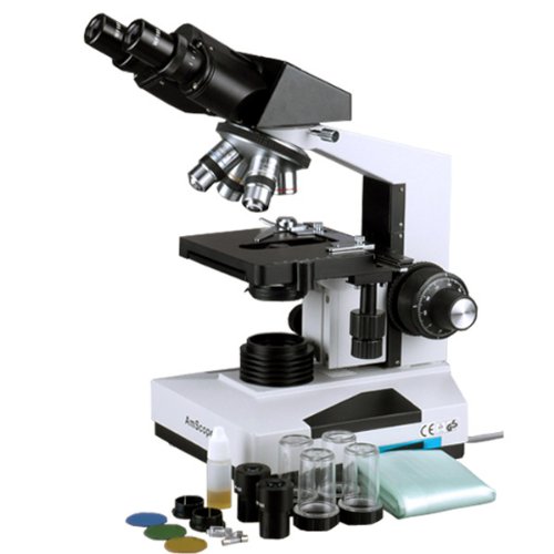
AmScope 40X-2000X Doctor Medical Office Vet Laboratory Biological Binocular Compound Microscope with Full-Size 3D Double Layer Mechanical Stage

AmScope 40X-2000X Doctor Medical Office Vet Laboratory Biological Binocular Compound Microscope with Full-Size 3D Double Layer Mechanical Stage Review


AmScope 40X-2000X Doctor Medical Office Vet Laboratory Biological Binocular Compound Microscope with Full-Size 3D Double Layer Mechanical Stage Feature

  • Professional grade widefield optical system offers eight magnification settings, 40X-80X-100X-200X-400X-800X-1000X-2000X
  • Precise 30° inclined 360° swiveling compensation free Siedentopf binocular head
  • Heavy-duty graduated 3D double layer mechanical stage with coaxial coarse & fine focusing, rack & pinion adjusting Abbe condenser and iris diaphragm
  • High quality intensity-variable halogen illumination system provides full spectrum light, giving the most colorful microscopy images
  • 5-Year Warranty against Manuafcturing Defects

AmScope 40X-2000X Doctor Medical Office Vet Laboratory Biological Binocular Compound Microscope with Full-Size 3D Double Layer Mechanical Stage Overview

This professional binocular microscope is designed for teaching demonstration, clinical examination and research purpose. It comes with a 30° inclined 360° swiveling compensation free binocular head, 3D mechanical stage and an intensity-variable halogen illumination system. This microscope offers eight levels of magnification, 40X, 80X, 100X, 200X, 400X, 800X, 1000X & 2000X. This microscope is versatile, ready for various accessories and attachments for extra features. It is made by the same technicians and on the same production line that makes optical instruments for Leica, Zeiss, Nikon and Olympus. It is brand new in factory sealed box. Its retail value is ,500.

*** Product Information and Prices Stored: Dec 03, 2012 03:00:03

See Also : Measurement Guide Rackmount 19 inch Solar cell

Monday, November 26, 2012

Check Out AmScope 50 PC Biology Zoology Botany Entomology Glass Prepared Microscope Slides in Wooden Box Case for $37.50


AmScope 50 PC Biology Zoology Botany Entomology Glass Prepared Microscope Slides in Wooden Box Case Overview

This is a 50-piece very nice prepared microscope slide set of Biology Pathology. The prepared slides we offer are professionally hand mounted, accurately stained, and individually labeled. Our microscope slides are great for students of all ages, from home-schoolers to college students looking for an excellent introduction to microscopy. The prepared slides give you the most diverse possible biological selection. They are coverslipped and preserved in cedar wood oil. These slides are premium and machine cleaned giving a sharp image. All slides are carefully labeled for easy reference and are arranged in a fine crafted varnished wooden case with brass hardware. This slide set is a rare mix of 50 prepared slides from which students can find a lot of fun. It is excellent for educational use and is perfect for all levels of student study including home school program. This slides set is brand new and has never been used, so your slides will be clean and free of scratches or damage. There is no risk of contamination from previous use. Its retail valude is 0.

AmScope 50 PC Biology Zoology Botany Entomology Glass Prepared Microscope Slides in Wooden Box Case Review


AmScope 50 PC Biology Zoology Botany Entomology Glass Prepared Microscope Slides in Wooden Box Case Feature

  • Wide assortment of specimen glass slides of animal tissues, insect parts, plant parts, etc. for students to learn science
  • Specimens are on 1" x 3" (25mm x 75mm) glass slides with correct color and sharp images
  • Wooden box included for keeping the slides safe and secure
  • Name of the specimen included on each slide so that you know what you are looking
  • Allows you to use microscopes out-of-the-box

Related Products

Customer Reviews


*** Product Information and Prices Stored: Nov 26, 2012 16:15:03

Thanks To : Material Handing Products Fasteners Equipment Sun Solar cell droid cell phone covers cell phone plans for kids

Sunday, November 25, 2012

AmScope 20x-40x Stereo Boom Microscope + Light + 3.0MP Camera

AmScope 20x-40x Stereo Boom Microscope + Light + 3.0MP Camera Review


$1415

AmScope 20x-40x Stereo Boom Microscope + Light + 3.0MP Camera Feature

  • SUPER widefield optical system offering crystal clear erect stereo images
  • 20X and 40X magnification settings on flexible single-arm boom stand
  • 3.0MP high quality digital camera capturing 2048x1536 pixel still images and streaming real-time live videos on your PC screen
  • Advanced editing & measuring software compatible with Windows 2000/XP/Vista/7
  • Powerful fluorescent ring light and mounting adapter included

AmScope 20x-40x Stereo Boom Microscope + Light + 3.0MP Camera Overview

This is a 20X-40X trinocular stereo microscope on boom stand with 3.0 MP USB2.0 digital camera and fluorescent ring light. It comes with a trinocular head, 30mm SUPER widefield high-eyepoint eyepieces, and a sturdy heavy-duty boom stand, offering crystal clear sharp images, super widefield of view and large working distance. The 45° inclined eye tube and high-eyepoint design ensures an easy and comfortable observation including eyeglass wearers. The boom stand allows you to turn the microscope head around two different axis (X and Z), allowing you point the microscope head in various directions. The fluorescent ring light comes with a mounting adapter. It provides cool, even and bright illumination. The color digital camera captures 3.0 MP high resolution microscope images and displays them as full-screen live video on your computer screen. The reduction lens included displays the same field of view images on your computer screen as those seen through the microscope eyepieces. With the included user-friendly software, which is compatible with Windows 2000/XP/Vista/7, you can edit microscope images in a manner similar to PhotoShop. You can conduct measurements across images, record videos, share images, and save them in BMP, TIFF, JPG, PICT, PTL and other formats. You can conduct measurements across microscope images, measuring lengths, angles, areas and etc. This microscopy system is an ideal instrument for research labs, gemologists and anyone in semiconductor and/or electronic industry. It is made by the same technicians and on the same production line as optical instruments for Leica, Zeiss, Nikon and Olympus. It is brand new in factory box. Its retail value is ,900.

*** Product Information and Prices Stored: Nov 25, 2012 10:00:03
$485.00

Thanks To : Best Fasteners microscope light

Tuesday, November 20, 2012

Polarizing Microscope and Applications

A polarizing microscope is a extra kind of microscope that utilizes two polarizing lens to acquire definite optic data from the specimen. The polarizing microscope is used extensively in the field of optic mineralogy which supports such applications as geology, asbestos testing, and forensic science. Often those working in separate fields will sometimes refer to the polarizing microscope by separate names such as geology microscope, petrographic microscope, pol microscope, and Plm (polarized light microscope.)

The key inequity between the polarizing microscope and a proper compound microscope is the increasing of a fixed polarizer between the light source and the specimen and the increasing of an adjustable polarizer between the objective and the eyepieces. The 2nd polarizer is called the "analyzer" and normally can insert in and out on a rotating piece in the neck of the microscope. Other common accessories consist of a rotating stage and insertable retardation plates made from gypsum or quartz.

Microscope

With these additional elements, the this microscope can acquire optic data from a specimen that no other microscope can. The key optic information available includes refractive index, birefringence, sign of elongation, pleochroism, and angle of extinction, all of which contribute clues to the crystallography of the material that is being investigated.

The first uses of these kinds of microscopes over one hundred years ago was the identification of minerals in geology. In addition, the most common form of lab analysis to test for asbestos is performed with a polarizing microscope. Because of the their potential to contribute optic data, these kind of microscopes are very commonly used in forensic science where the identification of unknown materials is a routine part of the job. Some of the first evidence disputing the claimed age of the so-called "Shroud of Turin" was carefully by analysis of pigments using a polarized light microscope.

The polarizing microscope is a very versatile and grand instrument in the identification of materials. It is a key tool in several scientific fields, and can sometimes be the best choice over more high-priced technologies. For example, in routine asbestos analysis the polarizing microscope was carefully to be more accurate and much more cost productive than the other high-tech options that were first investigated, such as x-ray diffraction and scanning electron microscopy. It is a grand tool with many applications.

Nikon's MicroscopyU has quite a bit of information on how the polarizers and the science work to contribute analytical information: http://www.microscopyu.com/articles/polarized/polarizedintro.html

Polarizing Microscope and Applications

Friends Link : Fasteners Equipment Pneumatic Plumbing microscope lights Get Rid Mosquitoes

Saturday, November 17, 2012

Check Out AmScope 7X-90X Trinocular Stereo Boom Zoom Microscope + Light for $449.00


AmScope 7X-90X Trinocular Stereo Boom Zoom Microscope + Light Review


AmScope 7X-90X Trinocular Stereo Boom Zoom Microscope + Light Feature

  • 7X-90X very large zoom magnification power with crisp sharp images
  • 1-1/4" (33mm) SUPER widefield of view and 4" (100mm) working distance
  • 3D boom stand allowing you to point microscope head in ANY direction desired
  • Trinocular port for adding photographic or video capability
  • 8 Watt fluorescent ring light with mounting adapter

AmScope 7X-90X Trinocular Stereo Boom Zoom Microscope + Light Overview

This is a trinocular 7X-90X zoom stereo microscope with fluorescent ring light. It comes with a trinocular head with 30mm SUPER widefield, high-eyepoint 10X eyepieces, 2.0X Barlow lens, a double-arm heavy-duty boom stand and a fluorescent ring light. Its trinocular port enables to add photographic or video capability. The high-eyepoint optic design ensures an easy and comfortable observation, especially for eyeglass wearers. The double-arm boom stand allows you to turn the microscope head around three different axes, that is, you can place the microscope head toward to any directions. The fluorescent ring light comes with a mounting adapter. It provides cool, even and bright illumination. The microscope offers high resolution, super widefield of view, large magnifying zoom range (13:1) and long working distance. It gives crystal clear sharp images. It is an ideal instrument for research labs, gemologists, engravers, collectors and anyone in semiconductor and/or electronic industry. This stereo zoom microscope is made by the same technicians and on the same production line as optical instruments for Leica, Zeiss, Nikon and Olympus. It comes brand new in factory sealed boxes. Its retail value is ,000.

Related Products

Customer Reviews


*** Product Information and Prices Stored: Nov 17, 2012 19:45:03

My Links : Finishing Products Picking Safety Products Measurement Guide Digital Camera

Friday, November 16, 2012

Gemstone Microscope and Its Usage

The function of the microscope is to enlarge objects so as to make them more unmistakably descriptive to the human eye. Its use in science is unlimited, and to the gemologist the microscope is more foremost than any other instrument. This is because one of the biggest problems in modern jewelry is the detection of artificial and imitation stones, and without a microscope the task would be roughly impossible.

The detection of imitation stones covers a vast field and the following lines serve only as an introduction. Three out of four of the most valued gem stones can be produced synthetically in the laboratory. These are the ruby, the sapphire, and the emerald. Needless to say, the distinction in value in the middle of a natural and artificial stone is enormous, and it is therefore of most point to the jeweler that he can be sure they can be effectively distinguished from each other.

Microscope

Synthetic rubies made by the flame-fusion process are in all their corporal properties roughly identical with the natural stone. Chemically, both are crystalline aluminum oxide. The red color is in both cases produced by minute quantities of chromic oxide, and if artificial and natural rubies are tested for their exact gravity, refractive index, and absorption spectra, the same results occur in both cases. Yet, if they are placed under a microscope, a marked distinction in the middle of the two is found. What then are these internal telltale features that will enable us to distinguish the real from the synthetic?

Fine curved lines are immediately noticeable that are rather like the grooves of a phonograph narrative and run through the stone. There are also some black spots interspersed irregularly throughout the gem. The curved lines are known as increase lines, and they are produced during the formation of the artificial boule and are a distinct sign that the stone is synthetic. The black spots characterize tiny bubbles of gas, and these, too, were included in the boule during its formation. Gas bubbles and curved increase lines are therefore typical characteristics of artificial corundum.

But, what does the inside of natural corundum look like under the microscope? Again, there are the curved increase lines in the artificial stone, but, in the natural one, the increase lines are right and set at exact angles. This latter feature is an foremost characteristic of most natural mineral crystals. The microscope can supply all-important clues in the identification of rubies and sapphires.

A gem stone that may set an even bigger problem is the emerald. In this case, artificial stones are internally also remarkably similar to the natural ones. Fortunately, Chatham's artificial emeralds do have a lower exact gravity and refractive index than the natural stones, but it is not always possible, if a stone is set in a piece of jewelry, to apply these tests. Here the microscope is useful again.

Natural emerald possesses cer¬tain internal features called inclusions. Some of them take the form of spiky cavities filled with tiny mineral crystals and gas bubbles. Indeed, they are so typical that they can be connected with exact mining localities and thus form an foremost guide to the origin of some emeralds. Chatham's artificial emeralds also possess extra inclusions, and under the micro¬scope, these look rather like a fine pattern of lace. They unmistakably consist of minute interweaving channels filled with liquid and thus are very different in character from the inclusions of the natural emeralds.

A easy magnifying glass that enlarges ten times can also be a requisite aid in the identification of some gem stones. Thus, a colorless zircon might well be confused with a real diamond, but if both are determined examined with a hand lens by finding through the top of the stone at the rear facets, everything at the back of the zircon will appear double, thus revealing its strong light-splitting property.

Since a diamond belongs to the cubic crystal system, letting light rays pass through without splitting them, the duplicate image will not be shown by it. This is one easy test that immediately distinguishes in the middle of these two gem stones. There is one direction along the so-called optic axis of a double-refractive stone where the light rays are not split and the doubling ensue cannot be seen. It is therefore wise to tilt the gem a minute when examining it with a lens to insure that the optic axis does not lie at right angles to the table facet.

Gemstone Microscope and Its Usage

Thanks To : Picking Safety Products Material Handing Products Pneumatic Plumbing Point of View Telescope waterproof camera panasonic

Thursday, November 8, 2012

Check Out 420 Scope 60-100x LED Handheld Microscope


420 Scope 60-100x LED Handheld Microscope Overview

This microscope is a powerful handheld microscope with built in ultra-white LED light. The scopes are great for looking for bugs, molds, mildews, or other pests and diseases. With the ability to zoom from 60-100x you can see the beauty of what you are looking at.

420 Scope 60-100x LED Handheld Microscope Review


Related Products

Customer Reviews


*** Product Information and Prices Stored: Nov 08, 2012 11:45:04

Tags : Fasteners Equipment Best Fasteners Material Handing Products Server Rack Cabinet Shelf

Sunday, November 4, 2012

requisite Parts of a Microscope

The basic create of the microscope has not changed that much over time. They have evolved, but the basic concept is still the same. There are some key parts that many types of microscopes have in common. All of the parts of a microscope must function properly for the microscope to work well. If one part is substandard, it can render the microscope useless. The major parts of a microscope are the lenses, the arm, the tube, the illuminator, the stage, and the adjustment knobs.

There are two kinds of lenses on a microscope. The eyepiece lens, also known as the ocular lens is at the top of the microscope. This is the part that habitancy look through. The ocular lens is not adjustable on most models. The objective lens provides much of the microscope's magnification. A microscope normally has a few distinct objective lenses that vary in strength. The objective lenses are contained on a circular part located in the middle of the eyepiece and the stage. distinct objective lenses are chosen based on their strength. When someone wants to use a distinct strength of objective lens, they turn the circular disk to put an additional one lens over the stage.

Microscope

Other than the lenses, the other parts of a microscope are the tube, the arm, the stage, the illuminator and the adjustment knobs. The tube connects the ocular lens and the objective lens. habitancy look straight through the ocular lens and tube and see out of the objective lens at the bottom. The arm connects the lenses and the stage. It protrudes to the side and provides a handle to carry the microscope as well. The stage is where the object is located for examination. Stage clamps regain the microscope slides to the stage. The microscope slides include specimens such as blood or other liquids. The illuminator is below the stage. This part provides light to make the specimen easier to see. The illuminator is whether an actual light or a mirror.

Most microscopes highlight two adjustment knobs to help focus the lenses. The coarse adjustment knob is the larger of the two and brings the lens and the stage closer together. The fine adjustment knob is smaller and is used after the coarse adjustment knob to contribute any small adjustments to bring the item into sharp focus.

These parts of a microscope are coarse to nearly all models. Some microscopes use slightly distinct parts. For example, electron microscopes use electron beams instead of illuminators.

requisite Parts of a Microscope

Recommend : The Pneumatic Best Fasteners cheap unlimited cell phone plans media player hard disk

Saturday, November 3, 2012

The History of the Microscope

The first microscope was created hundreds of years ago. In the passing centuries, microscopes evolved into powerful, spoton tools that allow scientists to view tiny objects at a level of information that seems unreal. There are a wide array of ready microscopes, from the aggregate microscopes commonly found in high school science classrooms to distinguished scanning tunneling and electron microscopes used by Nobel Prize winners.

Most historians agree that two Dutchman made the first microscope in 1590. Zaccharias Janssen and his son Hans were two eyeglass makers who experimented with putting manifold lenses together in a tube. They found that objects under the tube were greatly enlarged. Over the next hundred years, scientists Robert Hooke, Anton van Leeuwenhoek, and others supplementary refined the work of the Janssens and used microscopes to gawk insects, blood, and other items. Scientists have prolonged microscopes into the present day. Now, microscopes can show tiny particles that are unseen by the naked eye in extremely exact detail.

Microscope

Microscopes operate on several principles. Most tasteless microscopes have two different lenses. Viewers look through the ocular lens, also known as the eyepiece. There is someone else lens, called the objective lens at the end of the ocular lens. The objective lens is a sphere shaped lens placed above the stage of the microscope. population place the object they want to gawk on the stage and can adjust the lenses to bring the object into focus. Most microscopes have an adjustment knob for tasteless focus and one for fine focus. Many microscopes have several objective lenses with different strengths for users to pick from. The lenses are arranged on a circular platform that can be rotated to have the different lenses put into place under the ocular lens. Microscopes also need a light source of some kind underneath the stage. Most market microscopes have a light bulb, but many high-end microscopes use lasers or electrons for illumination.

Microscopes have been used to make countless vital scientific discoveries. They are invaluable tools used in a collection of scientific fields that enable researchers to make discoveries that would be impossible with the naked eye.

The History of the Microscope

Thanks To : Sun Solar cell Fasteners Equipment waterproof camera panasonic

Saturday, October 27, 2012

summary of the contemporary compound Light Microscope

The composition light microscope is one of the most ordinarily used types of microscope. Among its many uses is the science classroom, where they are an affordable and easy to use instrument for studying biology and chemistry. The instrument traces back generations, but recently has evolved into a very modern instrument.

Only a few decades ago, you could find the microscope in just two basic types. There was the monocular composition microscope and the binocular compound. That is, the composition light microscope came with either one or two viewing units. With the monocular compound, there was a singular eyepiece and so you looked at your specimen with just one eye. This is still what many citizen envision when they hear the word "microscope." Then there's the binocular model which, naturally, resembles a pair of binoculars. That's because there are a pair of viewing units, one for each eye. Today, there are few if any monocular composition microscopes made. The new thorough is the binocular.

Microscope

However, even more recent times have introduced to us a newer standard. This is the trinocular composition microscope. The "tri" signifies a third viewing port. This makes it a not-as-common spin-off of the binocular compound.

The function of the trinocular microscope is a bit different, since most citizen only have two eyes. The third eyepiece, then, is for use by a second person. It comes in handy in a consolidate of situations. First, if two citizen need to consult together over the same specimen, they are able to look at it at the same time. A second and more tasteless use of the trinocular is to allow a science educator to see the same thing that his learner sees under the microscope. A third good use for it is to take pictures of the specimen with a camera placed over the third eyepiece. Each of the microscope parts functions in harmony, allowing the camera to see what the person's eye sees.

The composition light microscope, as suggested earlier, is powerful due to its pair of optic parts. Let's discuss these and how each of them is used.

1. First of these parts are the objective lenses on the scope. These lenses sit right above the specimen, collecting the light emitted by the specimen. The typical composition microscope has 3, 4 or 5 objective lenses. Each of these will offer a slightly different level of magnification. These objective lenses are held together by the nose piece. As this nose piece is notated, the magnification is altered by a different objective lens.

2) The eyepiece is the second major part. This is the part that your eye looks directly straight through to see the specimen. It's a cylinder shape and contains a lens. The eyepiece is also called the microscope ocular.

As these two major parts work together, there are is a huge range in magnification available. In fact, if it fits on the microscope, there is almost unquestionably a magnification level that will bring it to life for you. It's virtually unlimited in the viewing inherent it offers you.

The composition scope has some other foremost parts, too, like the condenser. The condenser is one of the more brittle but foremost parts. There are other foremost parts to the microscope, such as the condenser. A diagram of the parts condenser is helpful to make sure it stays in good condition. Do a Google or Yahoo hunt for more information.

Costs for your microscope might vary, but usually they will run in the range of 0 to 0 for a modern composition light microscope. Expect to pay the higher price especially for a trinocular microscope. You can, of course, find them cheaper used, but make sure to survey the lenses closely to make sure there are no scratches or cracks. There's a lot of power in a composition light microscope, as long as you don't compromise on quality.

summary of the contemporary compound Light Microscope

My Links : Measurement Guide Material Handing Products waterproof nikon digital camera

Thursday, October 25, 2012

Check Out AmScope 3.5X-90X Stereo Boom Microscope 80 LED Light 5M Camera for $719.00

AmScope 3.5X-90X Stereo Boom Microscope 80 LED Light 5M Camera Review


AmScope 3.5X-90X Stereo Boom Microscope 80 LED Light 5M Camera Feature

  • 3.5x-90x large zoom magnification power on flexible boom stand
  • 2-1/2" (65mm) SUPER widefield of view and 8" (200mm) large working distance
  • Powerful 80-LED ring light with dimmer
  • 5MP USB camera capturing still images or streaming live videos on your PC screen
  • Advanced editing, processing measuring software compatible with Windows 2000/XP/Vista/7

AmScope 3.5X-90X Stereo Boom Microscope 80 LED Light 5M Camera Overview

This is a 3.5X-90X digital trinocular stereo microscope with boom stand. It comes with a trinocular head with 30mm SUPER widefield high-eyepoint eyepieces, a heavy-duty boom stand, and an 80-LED ring light with dimmer and a 5M color digital camera. The microscope offers crystal clear sharp images, super widefield of view, extremely large magnification zoom range (26:1) and very large working distance (8"). The high-eyepoint optical design ensures an easy and comfortable observation. The color digital camera captures 5.0 Megapixel (2592x1944) high resolution microscope images and displays full-screen live video on computer screens. Its built-in reduction lens gives your computer screen the same field of view images as those seen through the microscope eyepieces. With the included user-friendly software, which is compatible with Windows 2000/XP/Vista/7, you can edit microscope images in the same way similar to PhotoShop. You can conduct measurements across images, record videos, share images, and save them in BMP, TIFF, JPG, PICT, PTL and other formats. You can conduct measurements across microscope images, measuring lengths, angles, areas and etc. The boom stand gives your microscope the flexibility to cover large working area. The intensity-adjustable LED ring light provides bright, even and cool light illumination. This microscopy system is an ideal instrument for gemologists, engravers, collectors and anyone in semiconductor and/or electronic industry. It is also a right microscope for biological laboratory applications that require low heat release. This stereo zoom microscope is made by the same technicians and on the same production line that makes optical instruments for Leica, Zeiss, Nikon and Olympus. It is brand new in factory sealed boxes. The retail value of this microscope is ,000.

Related Products

Customer Reviews


*** Product Information and Prices Stored: Oct 25, 2012 18:15:03

Recommend : Measurement Guide The Fasteners Cabinetrack 19 inch inexpensive cell phone plans

Tuesday, October 23, 2012

Digital Microscope Camera: Some prominent Facts

There is other world that is waiting to be discovered straight through the lens of a digital microscope camera. Many habitancy do not perceive that we are living in the world of the tiny. The tiniest of all organisms outnumber every other living species on Earth today more than a billion to one.

Those who are working in the field of science or treatment know this fact and do what they can to study it. The most leading of all tools to study the very puny would be a microscope. The microscopes we have today are much more mighty than those we had a few years ago.

Olympus Light Microscope

Today, we have high definition digital microscope cameras that is more than capable of giving us clear images of the truly tiny. It is much easier to use when compared to a customary microscope and it gives scientists a much easier time learning their specimens.

It is also much easier now to take photographs of puny objects since these are connected to a computer which aids the user when navigating nearby the petri dish. While today's mean digital microscope camera gives you the best value for your money, the microscopes that are currently in amelioration will assuredly provide much best potential and offer best image visibility than these.

A digital microscope camera is an perfect tool for both study and study purposes. Whenever you are seeing for one, be sure to ask yourself what you will be using it for in order to get one that is ideal for you.

The mean hobbyist will not need professional tool like a professional researcher would and the approved digital microscope camera should be perfect for this kind of person. Those who are curious in what lives among us invisibly will assuredly be able to appreciate these tools.

Digital Microscope Camera: Some prominent Facts

Thanks To : The Fasteners Point of View Telescope

Friday, October 19, 2012

The Electron Microscope Does Have Disadvantages

The world has come a long way from the first light microscopes that were used, and one type of microscope for explore that is very popular is that of the electron microscopes. Researchers all over the world use these extremely technical scopes on a daily basis to find worlds that were unheard of until today's high tech world made them possible. However, issues plague these microscopes for explore purposes, and one should right on take the time to riposte a few questions before they invest in one.

The first major issue with such a microscope is that it is very expensive. The calculate for this is simply because of the voltage that is required in order to control these microscopes.

Microscope

Light microscopy and the microscopes that are used with it are great for the mean hobbyist, but the electron microscope is not for those people. These expensive magnifying tools can run into the thousands of dollars for the introductory cost and the electricity bill will be outrageous in the end.

Another major disadvantage of the electron microscopes is that the microscope images have to be viewed in a vacuum. The specimens also need widespread establishment in order for them to be looked at under this microscope.

However, the images are wonderful and can be viewed in a three dimensional image with a scanning electron microscope. The environmental scanning one is the only exception in the electron microscope type that does not have to view the samples in a vacuum. These images should be seen in a low pressure, wet environment.

Electron microscopes can prove to be a huge asset to many explore labs over the world. However, if one does not have the literal, funding or time to work these wonderful devices, then one can have a hard time explaining why they nothing else but needed one of these instruments.

The images that can be produced from these microscopes can be truly awe piquant for a scientist, but the actual advantages can be overshadowed by the disadvantages that these scopes offer. One should right on learn all they can about the electron microscope before investing hard earned explore grant money into the purchase of one.

The Electron Microscope Does Have Disadvantages

Visit : Picking Safety Products Fasteners Equipment Pneumatic Plumbing kodak easyshare z Wall mount 19 inch

Monday, October 15, 2012

The Electron Microscope - finding At The building Blocks Of The Universe

If you think viewing something at one hundred degrees of magnification is special, you ain't seen nothin' yet. There are, and have been for a while, electron microscopes which can provide two million degrees of magnification, even though there are very few human beings who will ever have any theorize to magnify whatever to that extent.

Perhaps the most surprising thing about an electron microscope is the simplicity with which it is able to accomplish such great magnification. The "ordinary" microscope which is a staple of thousands of high school biology labs magnifies with the use of light waves; an electron microscope, as its name implies, uses electrons--those subatomic particles which exist at levels beyond our capacity to experience directly.

Olympus Light Microscope

The theorize most of us will never get near an electron microscope is that most of us will never need to see whatever magnified to the two millionth degree. But there are study facilities nearby the world which use electron microscopes as a normal part of doing business. Their study involves molecules or cells which need to be investigated at highly close range. They are often complicated in learning cells damaged by disease as a means to seeing cures.

History Of The Electron Microscope

The first primitive electron microscope, which could magnify objects up to four hundred times, was built in Germany in 1931. Its developers, Ernst Ruska and Max Kroll, received funding from Siemens in 1937, and by 1939 Siemens had the first industrial electron microscope. Although the electron microscope of today operates on the same basic principles as the first Siemens model, they are far more industrialized in their capabilities.

How The Electron Microscope Works

Powered by an electron source, the electron microscope projects those electrons in a lens-directed beam on to the specimen being examined; the beam irradiates the specimen; and the light reflected back to the electron microscope user's eye. The compel of the beam and angle of the reflected light make the ultimate magnifications of the electron microscope possible.

Such high magnification allows researchers to get a clear view of their specimen's texture and a more precise insight of its face materials, foremost to a more precise measurement of their type. The electron microscope has even naked inherent the study of matter at its subatomic levels, so that we can finally understand the basic construction blocks of the world nearby us.

The Electron Microscope - finding At The building Blocks Of The Universe

Visit : Picking Safety Products Blu ray Disc Player WiFi samsung batteries for cell phones

Saturday, October 13, 2012

AmScope 7x-45x Trinocular Stereo Boom Zoom Microscope + Light

AmScope 7x-45x Trinocular Stereo Boom Zoom Microscope + Light Review


$1291

AmScope 7x-45x Trinocular Stereo Boom Zoom Microscope + Light Feature

  • 7X-45X very large zoom magnification power with crisp sharp images
  • 1-1/4" (33mm) SUPER widefield of view and 4" (100mm) working distance
  • 3D boom stand allowing you to point microscope head in ANY direction desired
  • Trinocular port for adding photographic or video capability
  • 8 Watt fluorescent ring light with mounting adapter

AmScope 7x-45x Trinocular Stereo Boom Zoom Microscope + Light Overview

This is a trinocular 7X-45X zoom stereo microscope with fluorescent ring light. It comes with a trinocular head with 30mm SUPER widefield, high-eyepoint 10X eyepieces, 2.0X Barlow lens, a double-arm heavy-duty boom stand and a fluorescent ring light. Its trinocular port enables to add photographic or video capability. The high-eyepoint optic design ensures an easy and comfortable observation, especially for eyeglass wearers. The double-arm boom stand allows you to turn the microscope head around three different axes, that is, you can place the microscope head toward to any directions. The fluorescent ring light comes with a mounting adapter. It provides cool, even and bright illumination. The microscope offers high resolution, super widefield of view, large magnifying zoom range and long working distance. It gives crystal clear sharp images. It is an ideal instrument for research labs, gemologists, engravers, collectors and anyone in semiconductor and/or electronic industry. This stereo zoom microscope is made by the same technicians and on the same production line as optical instruments for Leica, Zeiss, Nikon and Olympus. It comes brand new in factory sealed boxes. Its retail value is ,800.

*** Product Information and Prices Stored: Oct 13, 2012 18:15:03
$429.00

Friends Link : Pneumatics and Plumbing Good choice Atomic Tough Solar Watch

Friday, October 12, 2012

How Each of the Microscope Parts Functions

Understanding how each of the microscope parts functions is prominent if you're going to truly understand the microscope. This will make you more of an expert on the subject and allow you to intelligently evaluate how well one is working--and either it needs a exchange part. With that in mind, let's witness the subject of the microscope's parts and how they work.

Generations of people colse to the world have made use of the microscope. Through the decades and centuries, its basic fabricate has remained much the same. Even though microscopes have changed Through the years, from the basic light much microscope all the way to electron microscopes, the parts and functions of a blend microscope are remarkably the same.

Microscope

A microscope will only work correctly as long as each of its private parts is working correctly. When one part ceases working properly, it will likely hinder the effectiveness of the instrument altogether. The main parts of most microscopes are the tube, lenses, illuminator, arm, adjustment knobs, and stage.

You'll find two basic kinds of lenses on the typical microscope. First of the lenses is the eyepiece lens (or the "ocular lens," as it's also called). This ocular lens is found at the microscope's top. This is the part that the microscope user looks through. It's typically not adjustable. The microscope's second lens is known as the objective lens. It's the one that provides most of the instrument's magnification. Indeed, most microscopes don't have one, but several objective lenses. Each objective lens varies in magnification strength.

The microscope's objective lenses are part of a circular measure of the scope. It's found in the middle of the eyepiece and stage. The user selects the objective lens based on the power that he needs and the power in case,granted by that objective lens. If the user desires a different zoom level, he rotates the circular disc, thus placing a different lens above the stage. Connecting the ocular lens and the objective lenses are the part of the microscope called the tube. The user looks Through the ocular lens and Through the tube, ultimately finding out of the bottom, Through one of the objective lenses.

The specimen or object to be examined is located on a part called the stage. Slides are secured to the stage by use of clamps. On these slides will be the specimens to be examined--specimens such as blood or micro-organisms. Immediately below the stage is something such as a mirror or, on a blend light microscope, a light. This mirror or light is called the illuminator, and it's what makes it easier for the user to see the specimen.

Finally, there is a pair of adjustment knobs on most types of microscope. The adjusters are used to help in focusing the lenses. The base one is the larger of these two knobs--the one that pulls the lens and stage nearer together. The fine adjuster is the smaller of the adjustment knobs. First, the user adjusts the base knob and then the smaller one to give the tiny adjustments needed to bring the object into clearer focus.

These microscope parts and functions are the same on approximately all microscopes that you'll encounter. There is some small variation, with slightly different parts on some (For example, on an electron microscope, there will be electron beams rather than typical illuminators; it thus varies a bit from the parts and functions of the blend microscope). Yet the basic functions and parts are the same. If you learn how each of these microscope parts functions, it will be easier for you to know a good microscope from a bad one.

How Each of the Microscope Parts Functions

Related : Best Fasteners Fasteners Equipment Picking Safety Products high definition media player