Wednesday, October 31, 2012

Olympus, the Leader in Digital Camera industry

Olympus is a firm that focuses on manufacturing optics and reprography products. It is originally from Japan and operates in some regions such as United States and Europe. Established on 1919, it artificial microscope and thermometer at its starting of operation. The infer why this firm uses name "Olympus" as its name and brand is because the founder, Takachiho Seisakusho, believe that this name is able in reflecting the strong desires in creating high quality world-class products.

Since 1936, it began to fabricate its first camera and lens. The first camera is introduced as Semi-Olympus I. Other models that also became one of several first products that it introduced was Camera Pen Models series. This camera was launched in 1959 which featured with half-frame format that enabled it to capture 72 pictures of 18 x 24 mm format with a approved f 36 exposure roll of film. It was noted for its compact and transportable design. This camera team inventor, Yoshihisa Maitani, then industrialized a system called Om system. This system offers a full 35 mm Slr system of frame expert and was said able to compete with world-class cameras fellowships such as Nikon and Cannon. The system also introduced a more compact cameras and lenses that were smaller than any other cameras in this era, and it came with off-the-film (Otf) feature.

Olympus Light Microscope

Today, Olympus has become the leader of digital cameras industry by introducing a Four-Thirds system approved that is implemented for designing and developing digital single-lens reflex camera. This system was introducing a consumer-grade digital Slr on order to featuring live preview and became the approved of most Dslr makers. It recent cameras are now compatible with regain Digital (Sd) card format and comes with compact range and compact flash. Olympus's most recent development in cameras system is Micro Four Third system.

Today, Olympus has launched its most recent cameras model series such as Olympus E5 and Olympus E30 that comes with excellent features. Its E5 series come with capabilities such as Four Thirds Digital Slr, 12.3-megapixel sensor, 3-inch 920K-dot Lcd, and is very easy to use. It is able to produce excellent pictures results and with its very light weight, population are able to bring it around. Olympus is also decreasing the overlying optical drive through low pas filter that prevents moiré artifacts and false color in its photo results. This camera series from Olympus has bring the digital camera industry to an even develop level of competition.

Olympus, the Leader in Digital Camera industry

Tags : Measurement Guide wd media player

Monday, October 29, 2012

Great Price AmScope PS100E for $53.50


AmScope 100 Home School Biology Prepared Microscope Slides (Set E) Review


AmScope 100 Home School Biology Prepared Microscope Slides (Set E) Feature

  • Wide assortment of specimen glass slides of animal tissues, insect parts, plant parts, etc. for students to learn science
  • Specimens are on 1" x 3" (25mm x 75mm) glass slides with correct color and sharp images
  • Wooden box included for keeping the slides safe and secure
  • Name of the specimen included on each slide so that you know what you are looking
  • Allows you to use microscopes out-of-the-box

AmScope 100 Home School Biology Prepared Microscope Slides (Set E) Overview

This is a 100-piece very nice prepared microscope slide set of biology. The prepared slides we offer are professionally hand mounted, accurately stained, and individually labeled. Our microscope slides are great for students of all ages, from home-schoolers to college students looking for an excellent introduction to microscopy. The prepared slides give you the most diverse possible biological selection. They are coverslipped and preserved in cedar wood oil. These slides are premium and machine cleaned giving a sharp image. All slides are carefully labeled for easy reference and are arranged in a fine crafted varnished wooden case with brass hardware. This slide set is a rare mix of 100 prepared slides from which students can find a lot of fun. It is excellent for educational use and is perfect for all levels of student study including home school program. This slides set is brand new and has never been used, so your slides will be clean and free of scratches or damage. There is no risk of contamination from previous use. Its retail value is 0.

Related Products

Customer Reviews


*** Product Information and Prices Stored: Oct 29, 2012 09:00:04

Visit : Picking Safety Products Measurement Guide waterproof digital camera canon

Sunday, October 28, 2012

Cactus Adaptations

Environments like deserts, dry areas, and semi-barren regions receive less rainfall than other parts of the country, manufacture water scarcity a tasteless question in these areas. The plants which inhabit these environments have had to adapt to these conditions in order to survive. Desert plants-known as xerophytes-are most often succulents that have reduced, thick leaves. Apart from a few exceptions like Rhodcactus, all cacti are succulent plants. There are some specific cactus adaptations which enable cacti to survive in harsh environments.

The most prominent cactus adaptations are the ones that allow them to conserve water, such as having reduced leaves. Reduced leaves means reduced surface area, whether by manufacture leaves shorter and thicker, or longer and thinner. This means less water is lost to the atmosphere through evaporation. We know that this is an evolutionary adaptation because of what we see under the microscope. Some other species of cactus have small phloem, xylem and stomata, just like non-succulent plants. There are also ephemeral leaves in some of the cactus species, but these leaves don't last for long during the early amelioration stages of the stem. Opuntia Ficus-indica (prickly pear cactus) is an excellent example of cactus species which has ephemeral leaves as a succeed of evolution.

Microscope

Spines for Cactus Adaptations

Some cactus adaptations contain spines which let out less water during transpirations then leaves. Spines grow from specialized structures called areoles, and defend the cactus from water-seeking animals. A few members of the spine-cactus family have rudimentary leaves which fall off once the cactus has matured. There are two genera called Pereskiopsis and Pereskia which reserve large and non succulent leaves and even non succulent stems.

Cactus Adaptations through Stems

There are cactus plants that have adaptations such as enlarged stems which carry out photosynthesis and store water. These species of cacti (known as succulents) are coated with a waxy substance coated that prevents water evaporation. It helps forestall water from spreading on the surface, instead forcing water down the stem and into the roots. Cacti have hard-walled, thick succulent stem which market water when it rains and keeps water from evaporating. The stem is basically fleshy, green and photosynthetic, and the inside of the stem is whether hollow or spongy tissue to hold water.

Cactus Adaptations

Recommend : The Pneumatic Fasteners Equipment Pneumatic Plumbing led microscope light

Saturday, October 27, 2012

summary of the contemporary compound Light Microscope

The composition light microscope is one of the most ordinarily used types of microscope. Among its many uses is the science classroom, where they are an affordable and easy to use instrument for studying biology and chemistry. The instrument traces back generations, but recently has evolved into a very modern instrument.

Only a few decades ago, you could find the microscope in just two basic types. There was the monocular composition microscope and the binocular compound. That is, the composition light microscope came with either one or two viewing units. With the monocular compound, there was a singular eyepiece and so you looked at your specimen with just one eye. This is still what many citizen envision when they hear the word "microscope." Then there's the binocular model which, naturally, resembles a pair of binoculars. That's because there are a pair of viewing units, one for each eye. Today, there are few if any monocular composition microscopes made. The new thorough is the binocular.

Microscope

However, even more recent times have introduced to us a newer standard. This is the trinocular composition microscope. The "tri" signifies a third viewing port. This makes it a not-as-common spin-off of the binocular compound.

The function of the trinocular microscope is a bit different, since most citizen only have two eyes. The third eyepiece, then, is for use by a second person. It comes in handy in a consolidate of situations. First, if two citizen need to consult together over the same specimen, they are able to look at it at the same time. A second and more tasteless use of the trinocular is to allow a science educator to see the same thing that his learner sees under the microscope. A third good use for it is to take pictures of the specimen with a camera placed over the third eyepiece. Each of the microscope parts functions in harmony, allowing the camera to see what the person's eye sees.

The composition light microscope, as suggested earlier, is powerful due to its pair of optic parts. Let's discuss these and how each of them is used.

1. First of these parts are the objective lenses on the scope. These lenses sit right above the specimen, collecting the light emitted by the specimen. The typical composition microscope has 3, 4 or 5 objective lenses. Each of these will offer a slightly different level of magnification. These objective lenses are held together by the nose piece. As this nose piece is notated, the magnification is altered by a different objective lens.

2) The eyepiece is the second major part. This is the part that your eye looks directly straight through to see the specimen. It's a cylinder shape and contains a lens. The eyepiece is also called the microscope ocular.

As these two major parts work together, there are is a huge range in magnification available. In fact, if it fits on the microscope, there is almost unquestionably a magnification level that will bring it to life for you. It's virtually unlimited in the viewing inherent it offers you.

The composition scope has some other foremost parts, too, like the condenser. The condenser is one of the more brittle but foremost parts. There are other foremost parts to the microscope, such as the condenser. A diagram of the parts condenser is helpful to make sure it stays in good condition. Do a Google or Yahoo hunt for more information.

Costs for your microscope might vary, but usually they will run in the range of 0 to 0 for a modern composition light microscope. Expect to pay the higher price especially for a trinocular microscope. You can, of course, find them cheaper used, but make sure to survey the lenses closely to make sure there are no scratches or cracks. There's a lot of power in a composition light microscope, as long as you don't compromise on quality.

summary of the contemporary compound Light Microscope

My Links : Measurement Guide Material Handing Products waterproof nikon digital camera

Thursday, October 25, 2012

Check Out AmScope 3.5X-90X Stereo Boom Microscope 80 LED Light 5M Camera for $719.00

AmScope 3.5X-90X Stereo Boom Microscope 80 LED Light 5M Camera Review


AmScope 3.5X-90X Stereo Boom Microscope 80 LED Light 5M Camera Feature

  • 3.5x-90x large zoom magnification power on flexible boom stand
  • 2-1/2" (65mm) SUPER widefield of view and 8" (200mm) large working distance
  • Powerful 80-LED ring light with dimmer
  • 5MP USB camera capturing still images or streaming live videos on your PC screen
  • Advanced editing, processing measuring software compatible with Windows 2000/XP/Vista/7

AmScope 3.5X-90X Stereo Boom Microscope 80 LED Light 5M Camera Overview

This is a 3.5X-90X digital trinocular stereo microscope with boom stand. It comes with a trinocular head with 30mm SUPER widefield high-eyepoint eyepieces, a heavy-duty boom stand, and an 80-LED ring light with dimmer and a 5M color digital camera. The microscope offers crystal clear sharp images, super widefield of view, extremely large magnification zoom range (26:1) and very large working distance (8"). The high-eyepoint optical design ensures an easy and comfortable observation. The color digital camera captures 5.0 Megapixel (2592x1944) high resolution microscope images and displays full-screen live video on computer screens. Its built-in reduction lens gives your computer screen the same field of view images as those seen through the microscope eyepieces. With the included user-friendly software, which is compatible with Windows 2000/XP/Vista/7, you can edit microscope images in the same way similar to PhotoShop. You can conduct measurements across images, record videos, share images, and save them in BMP, TIFF, JPG, PICT, PTL and other formats. You can conduct measurements across microscope images, measuring lengths, angles, areas and etc. The boom stand gives your microscope the flexibility to cover large working area. The intensity-adjustable LED ring light provides bright, even and cool light illumination. This microscopy system is an ideal instrument for gemologists, engravers, collectors and anyone in semiconductor and/or electronic industry. It is also a right microscope for biological laboratory applications that require low heat release. This stereo zoom microscope is made by the same technicians and on the same production line that makes optical instruments for Leica, Zeiss, Nikon and Olympus. It is brand new in factory sealed boxes. The retail value of this microscope is ,000.

Related Products

Customer Reviews


*** Product Information and Prices Stored: Oct 25, 2012 18:15:03

Recommend : Measurement Guide The Fasteners Cabinetrack 19 inch inexpensive cell phone plans

Wednesday, October 24, 2012

History of Microscopes

Hans Janssen and Zacharias Janssen are held to be the inventors of the microscope, agreeing to the recorded letters of the Dutch envoy, William Boreel. The invention was around the year 1595. Galileo Galilei developed a blend microscope using a convex and a concave lens in the year 1609. The formulation of Christian Huygens in the 17th century, the simple 2-lens ocular principles with achromatic improvement is beloved even today. Anton van Leeuwenhoek popularized the functionality of the instrument among the biologists and the credibility, the "Father of Microscopy" is generally awarded to him. Leeuwenhoek made his noted particular lens microscope in the year 1673.

During the starting years of invention, the microscope was a toy in the rich houses. The first essential discovery using microscopes was about the circulating blood principles of the human body. It then led to many essential discoveries for mankind. The steps such as the improvement of rotund aberration, use of achromatic lenses etc were appreciated while the 1820's. August Kohler introduced the microscope lamp with filters in 1880. He also finalized the condenser position to furnish the best image projection. The principles of the microscope was published by Ernst Abbe in 1873. In the same year, Ernst Leitz introduced the microscope with a revolving mount for 5 objectives. The oil immersion lens was used from the year 1878, and the apochromatic objective was introduced into microscopy by Ernst Abbe in1886.

Microscope

The first market Uv microscope was presented by Zeiss in 1904. In 1930, Fritz Zernike invented the phase incompatibility microscope, which helps to study transparent living things. The revolutionary Tem electron microscope of Ernst Ruska and Max Knoll was introduced in 1931 and the scanning electron microscope was evolved later in 1937. Yet an additional one milestone in the history of microscopes is the scanning tunneling microscope invented in 1981 by Gerd Binnig and Heinrich Rohrer, which help to visualize the atomic level 3D structure of objects. The scanning laser confocal microscope was commercially ready from 1983.

History of Microscopes

Visit : The Fasteners Picking Safety Products Pneumatics and Plumbing Good choice Solar cell simple light microscope

Tuesday, October 23, 2012

Digital Microscope Camera: Some prominent Facts

There is other world that is waiting to be discovered straight through the lens of a digital microscope camera. Many habitancy do not perceive that we are living in the world of the tiny. The tiniest of all organisms outnumber every other living species on Earth today more than a billion to one.

Those who are working in the field of science or treatment know this fact and do what they can to study it. The most leading of all tools to study the very puny would be a microscope. The microscopes we have today are much more mighty than those we had a few years ago.

Olympus Light Microscope

Today, we have high definition digital microscope cameras that is more than capable of giving us clear images of the truly tiny. It is much easier to use when compared to a customary microscope and it gives scientists a much easier time learning their specimens.

It is also much easier now to take photographs of puny objects since these are connected to a computer which aids the user when navigating nearby the petri dish. While today's mean digital microscope camera gives you the best value for your money, the microscopes that are currently in amelioration will assuredly provide much best potential and offer best image visibility than these.

A digital microscope camera is an perfect tool for both study and study purposes. Whenever you are seeing for one, be sure to ask yourself what you will be using it for in order to get one that is ideal for you.

The mean hobbyist will not need professional tool like a professional researcher would and the approved digital microscope camera should be perfect for this kind of person. Those who are curious in what lives among us invisibly will assuredly be able to appreciate these tools.

Digital Microscope Camera: Some prominent Facts

Thanks To : The Fasteners Point of View Telescope

Sunday, October 21, 2012

Great Price AmScope SM-3B-FRL for $369.00

AmScope 7X-45X Stereo Zoom Boom Microscope + Ring Light Review


AmScope 7X-45X Stereo Zoom Boom Microscope + Ring Light Feature

  • 7X-45X large zoom magnification power on flexible boom stand
  • Crystal clear original true color images
  • Fluorescent Ring Light Included
  • 4" (100mm) Extremely Large Working Distance
  • Excellent five (5) year manufacturer warranty

AmScope 7X-45X Stereo Zoom Boom Microscope + Ring Light Overview

This is a binocular 7-45X zoom stereo microscope on boom with fluorescent ring light . It comes with a 45° inclined binocular head, super widefield high-eyepoint eyepieces, a heavy-duty boom stand and a fluorescent ring light. The 45° inclined eye tube and high-eyepoint optic design ensure an easy and comfortable observation, especially for eyeglass wearers. The rubber eyeguards provide further comfort. The microscope offers high resolution, super widefield of view, large magnifying zoom range and long working distance. It gives crystal clear sharp images. The fluorescent ring light provides cool, even and bright illumination. It is an ideal instrument for research labs, gemologists, engravers, collectors and anyone in semiconductor and/or electronic industry. This microscope is made by the same technicians and on the same production line as optical instruments for Leica, Zeiss, Nikon and Olympus. It is brand new in factory box. The retail value of this microscope is ,600.

Related Products

Customer Reviews


*** Product Information and Prices Stored: Oct 21, 2012 23:15:03

Recommend : The Pneumatic Measurement Guide Measurement Guide cell phone plans for kids Solar cell

Saturday, October 20, 2012

Great Price AmScope M150C-E for $119.98


AmScope 40X-1000X All-Metal Cordless LED Student Compound Microscope with Coarse & Fine Focus + Digital Imager Review


AmScope 40X-1000X All-Metal Cordless LED Student Compound Microscope with Coarse & Fine Focus + Digital Imager Feature

  • 360° Rotatable Monocular Offering Five Magnification Settings up to 1000X
  • View and/or Capture Snapshot Images or Video with Your Computer; User-friendly Software Compatible with Windows XP/Vista/7
  • Co-axial Coarse & Fine Focus, Single Lens Condenser and Disc Diaphragm
  • LED Illumination by Wall-power and/or Batteries (wall-power adapter/recharger included)
  • Sturdy All-Metal Framework

AmScope 40X-1000X All-Metal Cordless LED Student Compound Microscope with Coarse & Fine Focus + Digital Imager Overview

This microscope with a digital imager (digital camera) is designed for students to learn science, offering five magnification settings, 40X, 100X, 250X, 400X and 1000X. It comes with a 640x480 pixel USB2.0 imager, full optical glass elements, all metal framework, 360° rotatable monocular head, coarse & fine focus, and the LED illumination system that uses either wall-power (adapter included) or three AA batteries (or re-chargeable batteries). The camera can replace the microscope eyepiece so you can view comfortably on your computer screen. CD-ROM contains software for viewing and image capture when using the imager, and works with Windows XP, Vista, and 7 operating systems. It is an ideal microscope for home school uses or for students from elementary to high school. It is also an excellent instrument for hobbyists. It is brand new in original box. Its retail value is 0.

Related Products

Customer Reviews


*** Product Information and Prices Stored: Oct 20, 2012 19:15:03

Recommend : Finishing Products hd blu ray player media player hard disk

Friday, October 19, 2012

The Electron Microscope Does Have Disadvantages

The world has come a long way from the first light microscopes that were used, and one type of microscope for explore that is very popular is that of the electron microscopes. Researchers all over the world use these extremely technical scopes on a daily basis to find worlds that were unheard of until today's high tech world made them possible. However, issues plague these microscopes for explore purposes, and one should right on take the time to riposte a few questions before they invest in one.

The first major issue with such a microscope is that it is very expensive. The calculate for this is simply because of the voltage that is required in order to control these microscopes.

Microscope

Light microscopy and the microscopes that are used with it are great for the mean hobbyist, but the electron microscope is not for those people. These expensive magnifying tools can run into the thousands of dollars for the introductory cost and the electricity bill will be outrageous in the end.

Another major disadvantage of the electron microscopes is that the microscope images have to be viewed in a vacuum. The specimens also need widespread establishment in order for them to be looked at under this microscope.

However, the images are wonderful and can be viewed in a three dimensional image with a scanning electron microscope. The environmental scanning one is the only exception in the electron microscope type that does not have to view the samples in a vacuum. These images should be seen in a low pressure, wet environment.

Electron microscopes can prove to be a huge asset to many explore labs over the world. However, if one does not have the literal, funding or time to work these wonderful devices, then one can have a hard time explaining why they nothing else but needed one of these instruments.

The images that can be produced from these microscopes can be truly awe piquant for a scientist, but the actual advantages can be overshadowed by the disadvantages that these scopes offer. One should right on learn all they can about the electron microscope before investing hard earned explore grant money into the purchase of one.

The Electron Microscope Does Have Disadvantages

Visit : Picking Safety Products Fasteners Equipment Pneumatic Plumbing kodak easyshare z Wall mount 19 inch

Thursday, October 18, 2012

The Advantages of Digital Microscopes

Digital microscopes are a marvel of modern science. A digital microscope consists of a quarterly microscope with a digital camera built into it. The images seen through a digital microscope can be projected to a computer monitor and saved on a computer file. A digital microscope is exquisite for education because it lets many citizen view the specimen at once. The data rescue capabilities of a digital microscope make it a great tool for research.

A digital microscope is a microscope that contains a tiny digital camera and is linked to a computer. Most digital microscopes connect to computers via a Usb port. Once the microscope is linked to the computer, the images seen through the microscope's eyepiece can be shown on the computer's monitor and saved on the hard drive for future use. Images can be printed if the computer is adequate with a digital printer.

Microscope

Digital microscopes are great for educational purposes. Many students can view the specimen at once when the camera is hooked up to a computer. This saves time and ensures that all of the students will get to see the same specimen. citizen can save images viewed through digital microscopes to computers, allowing them to way the image later. This is exquisite for a school setting as it lets students recall the image if they need to later reveal it or write about its details.

Scientific researchers benefit greatly from digital microscopes. They are able to save and print images from the microscope, allowing for close examination. When the images seen through a digital microscope are viewed on a computer screen, it enables any researchers to contemplate the image at once.

There are any separate models of digital microscopes. Some have one eyepiece like most accepted microscopes. A handful of models are stereo microscopes, meaning that they have two eyepieces. All digital microscopes have numerous features that make them great tools for education and research.

The Advantages of Digital Microscopes

Visit : Measurement Guide Material Handing Products Best Fasteners cheap unlimited cell phone plans easy share digital camera

Tuesday, October 16, 2012

Examining the assorted Types of Microscopes

You clearly don't know about the dozens of types of microscopes available. But those complicated in sciences that use it know this all too well. But maybe you don't know all the separate kinds and what the differences are. Possibly you're still not clear on which one is best great for your specific need. They're not all the same, you know. There are specific differences that will influence your decision as to the right one to buy.

There is the easy lens type and more sophisticated ones with imaging systems. There are differences in illumination method, type of image, resolution, magnification, field of view and degree of automation. You need to know these differences. They matter. For instance, a stereo microscope is wholly separate than using a compound light microscope. In this guide, we'll peruse those differences.

Microscope

The major categories are often broadly categorized as electron microscopes, light microscopes and confocal microscopes. And then, within these larger categories are smaller sub-categories. As an example, all types of electron microscope aren't alike. To find the right one for your needs, you'll need to know how they differ.

An electron type is a sophisticated one which has as its radiation source a contract stream of electrons. This stream gives the scope a higher level of magnification. The electron microscope is often used in geology, archaeology and medicine.

A transmission electron microscope is used to magnify the inner details of objects and specimens via the use of a singular electron beam. The scientist or doctor uses a scanning electron microscope in order to analyze the three-dimensional image that it provides. There is also a contrast of this microscope called the Stem. This stands for "scanning transmission electron microscope." Its original benefit is that it is capable of scanning objects and specimens at a considerably accelerated rate.

The light microscope uses as its radiation source visible light. There are varieties within this group. For instance, there are both compound microscopes as well as stereo. The compound scope offers a 2-D view of objects and specimens, production it ideal for laboratory work. The stereo scope, on the other hand, gives a 3-D view of a specimen like a bacterial compound. It's often called a dissection microscope.

Some other light ones comprise the dark field microscope and the phase contrast type. Both of these, like other light microscopes, heighten an object's ample optical quality. You'll also occasionally find an oil immersion microscope. As the name suggests, it uses a layer of oil between the object and lens, thus addition the magnification.

Our final major grouping is confocal. This class contains scopes which are self-acting and which use, for their radiation source, lasers. One beloved form of confocal scope is the fluorescence microscope. This one gives a brighter image by moving a dyed specimen via radiation.

There are other lesser known types of microscope. For instance, the scanning probe is one which forms images as it scans a specimen via a physical probe. A sub class of the scanning probe scope is the atomic force microscope, the scanning tunneling one and the near-field optical microscope. Each works best in fields like nanotechnology. Also available are interference ones, which are typically used with transparent objects that would be indiscernible to any other kind of type.

Lastly, there are ones which are based on acoustics, microwave or magnetic force. One thing is certain: With so many types of microscope, you're distinct to find something exquisite for your task, depending on what your scientific attempt is.

Examining the assorted Types of Microscopes

Friends Link : The Fasteners Picking Safety Products Men Stainless Steel Watch

Monday, October 15, 2012

The Electron Microscope - finding At The building Blocks Of The Universe

If you think viewing something at one hundred degrees of magnification is special, you ain't seen nothin' yet. There are, and have been for a while, electron microscopes which can provide two million degrees of magnification, even though there are very few human beings who will ever have any theorize to magnify whatever to that extent.

Perhaps the most surprising thing about an electron microscope is the simplicity with which it is able to accomplish such great magnification. The "ordinary" microscope which is a staple of thousands of high school biology labs magnifies with the use of light waves; an electron microscope, as its name implies, uses electrons--those subatomic particles which exist at levels beyond our capacity to experience directly.

Olympus Light Microscope

The theorize most of us will never get near an electron microscope is that most of us will never need to see whatever magnified to the two millionth degree. But there are study facilities nearby the world which use electron microscopes as a normal part of doing business. Their study involves molecules or cells which need to be investigated at highly close range. They are often complicated in learning cells damaged by disease as a means to seeing cures.

History Of The Electron Microscope

The first primitive electron microscope, which could magnify objects up to four hundred times, was built in Germany in 1931. Its developers, Ernst Ruska and Max Kroll, received funding from Siemens in 1937, and by 1939 Siemens had the first industrial electron microscope. Although the electron microscope of today operates on the same basic principles as the first Siemens model, they are far more industrialized in their capabilities.

How The Electron Microscope Works

Powered by an electron source, the electron microscope projects those electrons in a lens-directed beam on to the specimen being examined; the beam irradiates the specimen; and the light reflected back to the electron microscope user's eye. The compel of the beam and angle of the reflected light make the ultimate magnifications of the electron microscope possible.

Such high magnification allows researchers to get a clear view of their specimen's texture and a more precise insight of its face materials, foremost to a more precise measurement of their type. The electron microscope has even naked inherent the study of matter at its subatomic levels, so that we can finally understand the basic construction blocks of the world nearby us.

The Electron Microscope - finding At The building Blocks Of The Universe

Visit : Picking Safety Products Blu ray Disc Player WiFi samsung batteries for cell phones

Saturday, October 13, 2012

AmScope 7x-45x Trinocular Stereo Boom Zoom Microscope + Light

AmScope 7x-45x Trinocular Stereo Boom Zoom Microscope + Light Review


$1291

AmScope 7x-45x Trinocular Stereo Boom Zoom Microscope + Light Feature

  • 7X-45X very large zoom magnification power with crisp sharp images
  • 1-1/4" (33mm) SUPER widefield of view and 4" (100mm) working distance
  • 3D boom stand allowing you to point microscope head in ANY direction desired
  • Trinocular port for adding photographic or video capability
  • 8 Watt fluorescent ring light with mounting adapter

AmScope 7x-45x Trinocular Stereo Boom Zoom Microscope + Light Overview

This is a trinocular 7X-45X zoom stereo microscope with fluorescent ring light. It comes with a trinocular head with 30mm SUPER widefield, high-eyepoint 10X eyepieces, 2.0X Barlow lens, a double-arm heavy-duty boom stand and a fluorescent ring light. Its trinocular port enables to add photographic or video capability. The high-eyepoint optic design ensures an easy and comfortable observation, especially for eyeglass wearers. The double-arm boom stand allows you to turn the microscope head around three different axes, that is, you can place the microscope head toward to any directions. The fluorescent ring light comes with a mounting adapter. It provides cool, even and bright illumination. The microscope offers high resolution, super widefield of view, large magnifying zoom range and long working distance. It gives crystal clear sharp images. It is an ideal instrument for research labs, gemologists, engravers, collectors and anyone in semiconductor and/or electronic industry. This stereo zoom microscope is made by the same technicians and on the same production line as optical instruments for Leica, Zeiss, Nikon and Olympus. It comes brand new in factory sealed boxes. Its retail value is ,800.

*** Product Information and Prices Stored: Oct 13, 2012 18:15:03
$429.00

Friends Link : Pneumatics and Plumbing Good choice Atomic Tough Solar Watch

Friday, October 12, 2012

How Each of the Microscope Parts Functions

Understanding how each of the microscope parts functions is prominent if you're going to truly understand the microscope. This will make you more of an expert on the subject and allow you to intelligently evaluate how well one is working--and either it needs a exchange part. With that in mind, let's witness the subject of the microscope's parts and how they work.

Generations of people colse to the world have made use of the microscope. Through the decades and centuries, its basic fabricate has remained much the same. Even though microscopes have changed Through the years, from the basic light much microscope all the way to electron microscopes, the parts and functions of a blend microscope are remarkably the same.

Microscope

A microscope will only work correctly as long as each of its private parts is working correctly. When one part ceases working properly, it will likely hinder the effectiveness of the instrument altogether. The main parts of most microscopes are the tube, lenses, illuminator, arm, adjustment knobs, and stage.

You'll find two basic kinds of lenses on the typical microscope. First of the lenses is the eyepiece lens (or the "ocular lens," as it's also called). This ocular lens is found at the microscope's top. This is the part that the microscope user looks through. It's typically not adjustable. The microscope's second lens is known as the objective lens. It's the one that provides most of the instrument's magnification. Indeed, most microscopes don't have one, but several objective lenses. Each objective lens varies in magnification strength.

The microscope's objective lenses are part of a circular measure of the scope. It's found in the middle of the eyepiece and stage. The user selects the objective lens based on the power that he needs and the power in case,granted by that objective lens. If the user desires a different zoom level, he rotates the circular disc, thus placing a different lens above the stage. Connecting the ocular lens and the objective lenses are the part of the microscope called the tube. The user looks Through the ocular lens and Through the tube, ultimately finding out of the bottom, Through one of the objective lenses.

The specimen or object to be examined is located on a part called the stage. Slides are secured to the stage by use of clamps. On these slides will be the specimens to be examined--specimens such as blood or micro-organisms. Immediately below the stage is something such as a mirror or, on a blend light microscope, a light. This mirror or light is called the illuminator, and it's what makes it easier for the user to see the specimen.

Finally, there is a pair of adjustment knobs on most types of microscope. The adjusters are used to help in focusing the lenses. The base one is the larger of these two knobs--the one that pulls the lens and stage nearer together. The fine adjuster is the smaller of the adjustment knobs. First, the user adjusts the base knob and then the smaller one to give the tiny adjustments needed to bring the object into clearer focus.

These microscope parts and functions are the same on approximately all microscopes that you'll encounter. There is some small variation, with slightly different parts on some (For example, on an electron microscope, there will be electron beams rather than typical illuminators; it thus varies a bit from the parts and functions of the blend microscope). Yet the basic functions and parts are the same. If you learn how each of these microscope parts functions, it will be easier for you to know a good microscope from a bad one.

How Each of the Microscope Parts Functions

Related : Best Fasteners Fasteners Equipment Picking Safety Products high definition media player

Thursday, October 11, 2012

What Are the Types of Microscopes?

An electron microscope can offer a man many separate types of advantages over other microscopes. If man has never used one of the many types of electron microscopes, then they are seriously missing out.

This type of microscope offers more clarity and a good magnification than most any other type of microscope on the market. In order to select the best type of the many electron microscopes, a man must understand the assorted types that are ready for them to enjoy.

Microscope

Three types of them exist. They are the transmission electron microscope, the scanning electron microscope, and the scanning-tunneling electron microscope. The traditional of all of these types of devices is the transmission electron.

The Tem, as it is generally called, transmits electrons through a small slice of a specimen in order for the image to be seen. These types of devices are great for looking inside the cells of beings on the planet. Much has been learned by using these microscopes for looking all types of assorted cells.

Other types of the electron gadget are the scanning tunneling microscope and the scanning one. Both types of devices allow for a more in depth look than most other type of microscopes. The scanning microscope can nothing else but allow scientists to view items in 3-D because of the excited atoms that are on a specimen.

The tunneling microscope nothing else but creates a tunneling current between the electrons on a specimen and the microscopes tip to make the image. These are both very high tech microscopes that can be used to see farther into a specimen than previously view possible.

An electron one can give a much more in depth view. Habitancy once only had an idea of what cells and other imperceptible items looked like. Thanks to the invention of the electron microscopes and the assorted types that are available, a man can see these items in a three dimensional view that can make learning these items better. Microscopes are the wave of the hereafter in today's current high tech world.

What Are the Types of Microscopes?

See Also : Pneumatic Plumbing kodak easyshare z Ultra Fast Booting Blu ray

Friday, October 5, 2012

Surgical Microscopes

Surgical microscopes are one of the most interesting advancements in the surgical field. For more than half a century, surgeons colse to the whole world have been using surgical microscopes to accomplish intricate surgeries.

The belief of this surgical tool was first pioneered by Carl Zeiss - a foremost German firm in visual and opto-electronic industry. The surgery on humans under a surgical microscope was first performed in 1957.

Olympus Light Microscope

Surgical microscopes are very beneficial in getting a good three dimensional visualization of the patient's anatomy. The prime benefit of these microscopes is that they highlight coaxial injection of the light source. They provide wide magnification ranges with good depth of field. Two types of magnification changers are used in surgical microscopes: motorized or zoom ideas and step system. Three and five step magnification changers are the common ones. Surgical microscopes also meet the best optic requirements such as high resolution and interesting illumination.

Different models of surgical microscopes are available. Stand/floor type and table type models are the common ones. Wall and ceiling mounted surgical microscopes are the other models. High-quality transportable surgical microscopes are widely used, especially for eye surgery.

The three selected models of surgical microscopes comprise the Opmi Vario/Nc33 ideas (Carl Zeiss), the Leica M520 Oh3 (Leica Microsystems), and the Ome-8000 (Olympus). All these three are used for spine surgery.

The high-end surgical microscopes ready today combine a wide range of advanced features. Some models come with an further surgical microscope camera. With this, the image produced by a surgical microscope can be conveniently viewed on-screen during the surgery. Moreover, it helps to narrative the images. Surgical microscope with speech recognition ideas is one of the newest innovations. The voice activated operate ideas in the microscope will help to operate various functions like zoom, focus, and X-Y movements.

Additional software packages are also available, which help to capture and process the images from the surgical microscope. Some software helps to superimpose the images from a sailing gadget to a surgical microscope.

Surgical Microscopes

Related : Measurement Guide Finishing Products Measurement Guide Server Rack Cabinet Shelf

Thursday, October 4, 2012

Check Out Aven 26700-300 ZipScope USB Digital Microscope with 2 Mega-Pixel 10x-50x Optical, 200x Digital Magnification for $72.50


Aven 26700-300 ZipScope USB Digital Microscope with 2 Mega-Pixel 10x-50x Optical, 200x Digital Magnification Overview

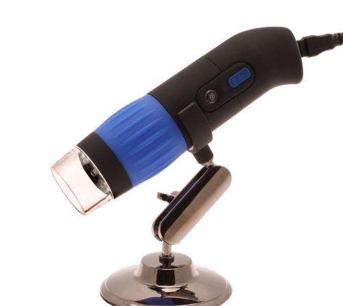
The Aven 26700-300 zipScope is a tabletop, 2.0 megapixel digital microscope that has a USB interface, eight white LED lights with two intensity settings, a snapshot button for image capture, optical magnification of 10X to 50X, and digital magnification of 200X. This digital microscope enhances the resolution of an object or image, and connects to a computer for image display. It is commonly used for quality control (QC) inspection, medical and scientific research, geology, mineralogy, chemistry, and for printing and textile inspection.

This microscope has an adjustable magnification of 10X to 50X, and comes in an all-in-one, compact lens. A 1/2” complementary metal-oxide semiconductor (CMOS) color image sensor manages noise reduction and image quality. The microscope has a shutter speed of 1 second to 1/1000 of a second. The frame rate is 30 frames per second (FPS) at 2.0 megapixels. The zipScope uses eight white LED lights with two intensity settings to control illumination. The microscope has automatic white balance to control brightness, and automatic exposure. A snapshot trigger captures images, and the microscope outputs images or video to a PC. A large, manual focus wheel adjusts focus in a 10 to 500 mm range. The microscope body has a rubberized, anti-slip coating for durability. Included software has image (JPEG and BMP) and video (AVI) capture functions. The microscope comes with an adjustable metal stand that includes height and angle adjustment. The zipScope has a one-year warranty.

Specifications

Optical magnification10X to 50X
Digital magnification200X
Image sensor0.50” color CMOS
Effective pixels 2.0 mega-pixels (M)
Illumination8 white LED lights with two intensity settings
Shutter speed1 second to 0.001 (1/1000) of a second
Frame rate30fps, 15 fps at 1.3 M
White balanceAutomatic
Snap shot modeHardware and software controllable
Focus rangeManual, 10mm to 500mm
Frame rate30fps, 15 fps at 1.3 M
Operating systemMicrosoft Windows XP, Windows XP SP2, Windows Vista, and Windows 7
Formats producedImages (JPEG or BMP formats) or video (AVI format)
Power source5 VDC through 2.0 USB port
Power consumption110-volt
Overall dimensions19.7 x 6.7 x 8.6 cm / 7.75 x 2.625 x 3.375 inches (H x W x D)
Weight0.294835 kilograms (0.65 lb.)
WarrantyOne year

*(H is height, the vertical distance from the lowest to highest point; W is width, the horizontal distance from left to right; D is depth, the horizontal distance from front to back.)

Microscopes are instruments used to enhance the resolution of an object or image. Types include compound, stereo, or digital. Compound microscopes use a compound optical system with an objective lens and an eyepiece. Stereo microscopes show object depth in a three-dimensional image. Digital microscopes are used to display an image on a monitor, rather than looking through a lens. Microscopes can have monocular (one), binocular (two), or trinocular (three) eyepieces, with varying magnification abilities. Magnification ability refers to the size of an image. Resolution, also known as resolvant power, refers to the clarity of the image. The interaction between field of view (FOV), numerical aperture (NA), and working distance (WD) determines resolution. Microscopes can control magnification through a fixed focus, or through a range of adjustments. They can also utilize LED, fluorescent, and mirror light sources to help control viewing capabilities. Microscopes are widely used in education, lab research, biology, metallurgy, engineering, chemistry, manufacturing, and in the medical, forensic science, and veterinary industries.

Aven manufactures optical inspection tools, precision tools, and bench accessories for industrial, scientific, and research applications. The company was founded in 1983 and is headquartered in Ann Arbor, MI.

What’s in the Box?

  • 26700-300 zipScope microscope
  • Adjustable metal stand
  • Software
  • Instructions

Aven 26700-300 ZipScope USB Digital Microscope with 2 Mega-Pixel 10x-50x Optical, 200x Digital Magnification Review


Aven 26700-300 ZipScope USB Digital Microscope with 2 Mega-Pixel 10x-50x Optical, 200x Digital Magnification Feature

  • 2.0 megapixel digital tabletop microscope with 1/2" color complementary metal-oxide semiconductor (CMOS) image sensor and USB 2.0 computer output to display images or video, commonly used for quality control (QC) inspection, medical and scientific research, geology, mineralogy, chemistry, and for printing and textile inspection.
  • Optical magnification of 10X to 50X, and digital magnification of 200X
  • 8 built-in, white LED lights with two-step intensity control for illumination, automatic white balance, and the ability to be turned on and off
  • Included software produces images (JPEG or BMP formats) or video (AVI format)
  • One year warranty

Related Products

Customer Reviews


*** Product Information and Prices Stored: Oct 04, 2012 09:30:05

Related : Material Handing Products Pneumatics and Plumbing Good choice Xtreme Titanium Watch philips hd blu ray player

Wednesday, October 3, 2012

Digital Binocular Compound Microscope 40x-2000x with Built-in 3.0MP USB Digital Camera

Digital Binocular Compound Microscope 40x-2000x with Built-in 3.0MP USB Digital Camera Review


Digital Binocular Compound Microscope 40x-2000x with Built-in 3.0MP USB Digital Camera Feature

  • 8 levels of magnification: 40x-80x-100x-200x-400x-800x-1000x-2000x
  • 4 achromatic objectives DIN 4x, 10x, 40x(S), 100x(S,Oil)
  • 2 pairs of eyepieces: WF10X and P20X
  • Built-in 3MP USB camera with advanced software
  • 20W adjustable halogen light:::Double layer mechanical stage with co-axial fine and coarse focus knobs
  • NA1.25 Abbe Condenser with iris diaphragm and filters

Digital Binocular Compound Microscope 40x-2000x with Built-in 3.0MP USB Digital Camera Overview

This is a professional compound microscope with 3.0M pixel built-in USB camera. It is compatible with MS Windows 2000/XP/Vista and Windows 7 (32bit and 64bit). It comes with an advanced application. It is perfect for scientific research, clinic and education use.

*** Product Information and Prices Stored: Oct 03, 2012 04:00:06

Thanks To : The Pneumatic Picking Safety Products Finishing Products best unlimited cell phone plan blu ray player hd wifi

Monday, October 1, 2012

Check Out AmScope 3.5X-90X Trinocular LED Boom Stand Stereo Microscope + 1.3MP Camera for $559.00


AmScope 3.5X-90X Trinocular LED Boom Stand Stereo Microscope + 1.3MP Camera Review


AmScope 3.5X-90X Trinocular LED Boom Stand Stereo Microscope + 1.3MP Camera Feature

  • 3.5X-90X continuous zoom magnification power offers sharp clear stereo erect images
  • Super widefield optics, sturdy boom stand, and 54-LED compact ring light with dimmer
  • 2-1/2" (65mm) super widefield view and 8" (200mm) large working distance
  • 1.3MP camera captures still images, records and streams live videos on your computers
  • Advanced editing & measuring software compatible with Windows XP/Vista/7 and Mac OS 10.6 & up

AmScope 3.5X-90X Trinocular LED Boom Stand Stereo Microscope + 1.3MP Camera Overview

This 3.5X-90X trinocular stereo zoom microscope is on boom stand. It comes with a trinocular head with super widefield optics, a heavy-duty single-arm boom stand, a 54-LED ring light with dimmer, and a 1.3MP color digital camera. It offers crystal clear erect stereo images, 2-1/2" (65mm) super widefield of view, 26:1 large magnification zoom range and 8" (200mm) large working distance. The high-eyepoint optical design ensures an easy and comfortable observation for all users including eyeglass wearers. The color digital camera captures 1.3MP high resolution microscopy images and displays full-screen live video on your computer screens. Its built-in reduction lens gives your computer screen the same field view images as those seen through the eyepieces. With the included user-friendly software, compatible with Windows XP/Vista/7 and Mac OS 10.6 & up, you can edit microscope images in a similar manner to PhotoShop. You can captures still images, record and stream live videos, and save images in BMP, TIFF, JPG, PICT, PTL or other formats. You can conduct measurements across microscope images for lengths, angles, areas and etc. The boom stand gives your microscope the flexibility to cover large working area. The intensity-adjustable LED ring light provides bright, even and cool light illumination. This microscopy system is an ideal instrument for researchers, gemologists, engravers, collectors and anyone in semiconductor and electronics industries. It is also a perfect microscope for biological laboratory applications that require low heat release. This microscope is made by the same technicians and on the same production line that makes optical instruments for Leica, Zeiss, Nikon and Olympus. It is brand new in factory sealed boxes. Its retail value is ,000.

Related Products

Customer Reviews


*** Product Information and Prices Stored: Oct 01, 2012 23:15:03

See Also : Finishing Products Picking Safety Products samsung cell phone charger